
[ad_1]
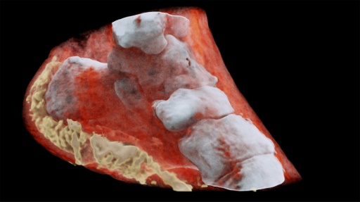
New Zealand scientists have made the first-ever three-dimensional (3D) color X-ray of a human body, using a technique that could help Improving medical diagnosis, according to the CERN European physics laboratory using the technology
The new device, based on traditional black-and-white radiography, incorporates the particle tracking technology developed for the large accelerator of CERN's Large Hadron Collider (LHC), which discovered the elusive elemental particle Higgs Boson in 2012.
"This color X-ray imaging technique could produce clearer images and more accurate and help doctors to give more accurate diagnoses to their patients, "says CERN in a statement.
According to CERN , the images clearly show the difference between bone, muscle and cartilage, but also the position and size of cancerous tumors, for example
CERN technology, called Medipix, works like a camera detecting and counting individual subatomic particles when they collide with pixels while their electronic shutter is open.
This allows high resolution and high contrast images.
Thus, this new imaging tool allows to obtain images that no other imaging device can achieve, according to developer Phil Butler of the University of Canterbury, New Zealand
The New Zealand company MARS Bioimaging Ltd, markets this 3D scanner , called "Spectral CT".
In the coming months, this scanner, equipped with a Medipix reader chip, will be the subject of a first clinical trial on patients in orthopedics and rheumatology. New Zealand, paving the way for a potentially routine use of this next-generation equipment, according to CERN.
Source link
