[ad_1]
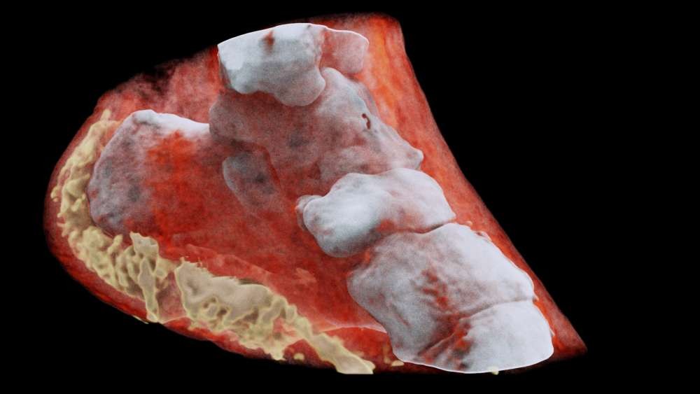
<img alt = "Detailed image of X-ray scanning (Photo: Disclosure)" height = "563" src = "https://s2.glbimg.com/lfXfPNNnF-E7_kECdxVIxkUdUo4=/e.glbimg.com / og / ed / X-ray scanning of the radiographic image of a radiographic image of a radiographic image of the radiographic image of an X-ray image. The study of subatomic particles never observed before by scientists is not the only function of the Large Hadron Collider developed by the European Organization for Nuclear Research (CERN) "The same technology for particle tracking will be applied in medicine and promises to revolutionize imaging diagnoses: using Medipix 3, a scanner created by CERN researchers can display color and three-dimensional images of bone tissue and structures in our body. compared to current X-ray machines.
In order to obtain the detailed images, Medipix 3 tracks electromagnetic radiation particles from the part of the body that will be badyzed. As a digital camera, the camera differentiates each particle and displays the details of the different "layers" of our body.
An algorithm that works near the device is responsible for interpreting all information received and converting them into images, which provide a detailed visualization with the actual layout of tissues, muscles and bones – including the density of grease, liquids and the concentration of minerals.
<img alt = "Device manages to detail the different structures of the human body (Photo: Divulgação)" height = "480" src = "https://s2.glbimg.com/ZHLUq3dpKi-H2oYnElHzDRfEZ_s=/e.glbimg .com / og / ed /f/original/2018/07/14/content-1531504531-wrist-mars-image-with-real-hand.jpg "title =" The device manages to detail the different structures of the human body (Photo: Divulgação) "width =" (19659002) Discovered in 1895 by German scientist Wilhelm Röntgen, X-rays are a form of electromagnetic radiation (just like light).
[ad_2]
Source link