[ad_1]
A team of researchers from University Medical Center (UMC) Utrecht in the Netherlands experiment with 3D bioprinted tissues that can be implanted into a living joint affected by arthritis [19659005]. The method should allow the replacement of damaged cartilage .
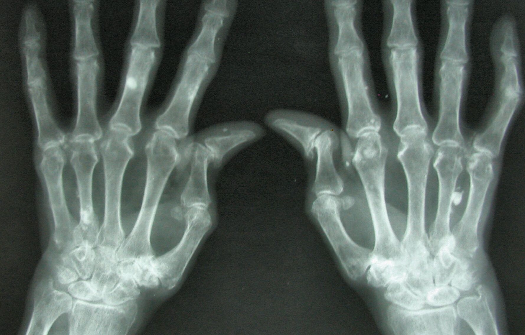
Arthritis is a common disease affecting approximately 350 million people worldwide. With more than 100 types of arthritis, this disease involves the disintegration of cartilage tissue in the joints, leading to pain, stiffness, and inflammation.
Using 3D bioprinting, patients can receive specific and on-demand cartilage as original cartilage to strengthen joints and reduce joint pain.
"Printing is not the last stage of biofabrication because printing something heart-shaped does not actually make a heart," said Jos Malda, professor of biofabrication in translational regenerative medicine at UMC Utrecht.
"Printed construction needs time and the correct chemical and biophysical indices to mature in a functional fabric."
Bioinks and Experimentation
As part of [19659002] Project 3D JOINT funded by the Horizon Vision 2020 of the EU [19659002] to encourage innovation in research on the continent, Professor Malda and his team rely on the capabilities of 3D printers and stem cell deposition.
Using the 3D bioprinting technique and following a precise medical plan, stem cells are deposited by 3D printers creating layer-by-layer complex tissues.
As demonstrated by Bioprinted Human Biota of the University of Newcastle Bioink Solutions Created from Stem Cells and Hydrogels of Donor ] – a clbad of materials made up of large molecules such as polymers – can produce living conditions for the organisms of the human body
. the challenge of maintaining the right conditions for a cellular building material, Professor Malda integrated hydrogel into the cartilaginous tissue.
"For bioprinting, the material must be able to keep the cells alive, which requires aqueous conditions and relatively low temperature processing, making hydrogel-based materials the ideal candidates. "
However, the softness of these hydrogels is at a disadvantage, according to Professor Malda. This is due to his inability to resist the mechanical forces that certain tissues undergo in the body.
"Strengthening the hydrogel makes it stronger – just as steel rods are combined with soft cement to create the reinforced concrete that makes the foundations of our homes," said the professor. Malda. His team uses a 3D printing technique called molten electro-writing that combines polycaprolactone fused, a type of polyester, with an electric field that creates fibers as fine as a hair to create a scaffold.
"The combination of the hydrogel with the fibers acts synergistically, increasing the composite's resistance more than 50-fold while allowing the cells to generate an extracellular matrix and mature into a cartilage-like tissue."
His team is We are now developing this process to create larger structures while including different materials for the replacement of combined bone and cartilage tissue. This will eventually lead to UMC Utrecht purpose of finally printing a complete joint in 3D
Also using 3D bioprinting technology is BIOLIFE4D a biotech company from Chicago, who successfully demonstrated his ability to 3D print human heart tissue.
Stay up-to-date with the latest in 3D printing by subscribing to bulletin 3D Printing Industry . Also, follow us on Twitter and like us on Facebook .
Looking for a change of pace or looking for new talent? Search and Publish 3D Print Jobs for opportunities and new talents across engineering, marketing, sales and more.
The featured image shows a hands arthritis X-ray. Image via Medical Xpress.
[ad_2]
Source link
