[ad_1]

A new sensor designed by MIT researchers could dramatically speed up the process of diagnosing sepsis, the leading cause of death in US hospitals, killing nearly 250,000 patients a year.
When time is running out in hospitals, an automated system can detect an early biomarker for a life-threatening disease.
Sepsis occurs when the body's immune response to infection triggers a chain reaction of inflammation throughout the body, resulting in high heart rate, high fever, shortness of breath, and other problems. If nothing is done, it can lead to septic shock, a drop in blood pressure and organ closure. To diagnose sepsis, doctors traditionally use various diagnostic tools, including vital signs, blood tests, as well as other imaging and laboratory tests.
In recent years, researchers have discovered protein biomarkers in the blood that are early indicators of sepsis. Interleukin-6 (IL-6), a protein produced in response to inflammation, is a promising candidate. In patients with sepsis, IL-6 levels may increase several hours before other symptoms occur. But even at these high levels, the concentration of this protein in the blood is generally too low for traditional baday devices to detect it quickly.
In an article presented this week at the Engineering in Medicine and Biology conference, MIT researchers describe a microfluidics-based system that automatically detects clinically significant IL-6 levels for the diagnosis of sepsis in approximately 25 minutes, using less than a blood stain. .
In a microfluidic channel, antibody microbeads mix with a blood sample to capture the biomarker of IL-6. In another channel, only the beads containing the biomarker bind to an electrode. A continuous voltage across the electrode produces an electrical signal for each bead containing a biomarker, which is then converted to a biomarker concentration level.
"For acute diseases, such as sepsis, which evolve very rapidly and can be life-threatening, it is useful to have a system that quickly measures these scarce biomarkers," says lead author Dan Wu, a PhD student in the department. mechanical engineering. "You can also frequently monitor the course of the disease."
Joel Voldman, Professor and Associate Director of the Department of Electrical and Computer Engineering, co-director of the Medical Electronic Devices Realization Center and Senior Research Scientist at the Electronics and Microsystems Labs, joined Wu.
Integrated and automated design
Traditional tests that detect protein biomarkers are large, expensive machines relegated to laboratories that require about one milliliter of blood and produce results within hours. In recent years, "point-of-care" portable systems have been developed that use microliters of blood to achieve similar results in about 30 minutes.
But point-of-care systems can be very expensive because most use expensive optical components to detect biomarkers. In addition, they only catch a small number of proteins, many of which are some of the most abundant in the blood. Any effort to reduce the price, reduce the components or increase the protein ranges has a negative impact on their sensitivity.
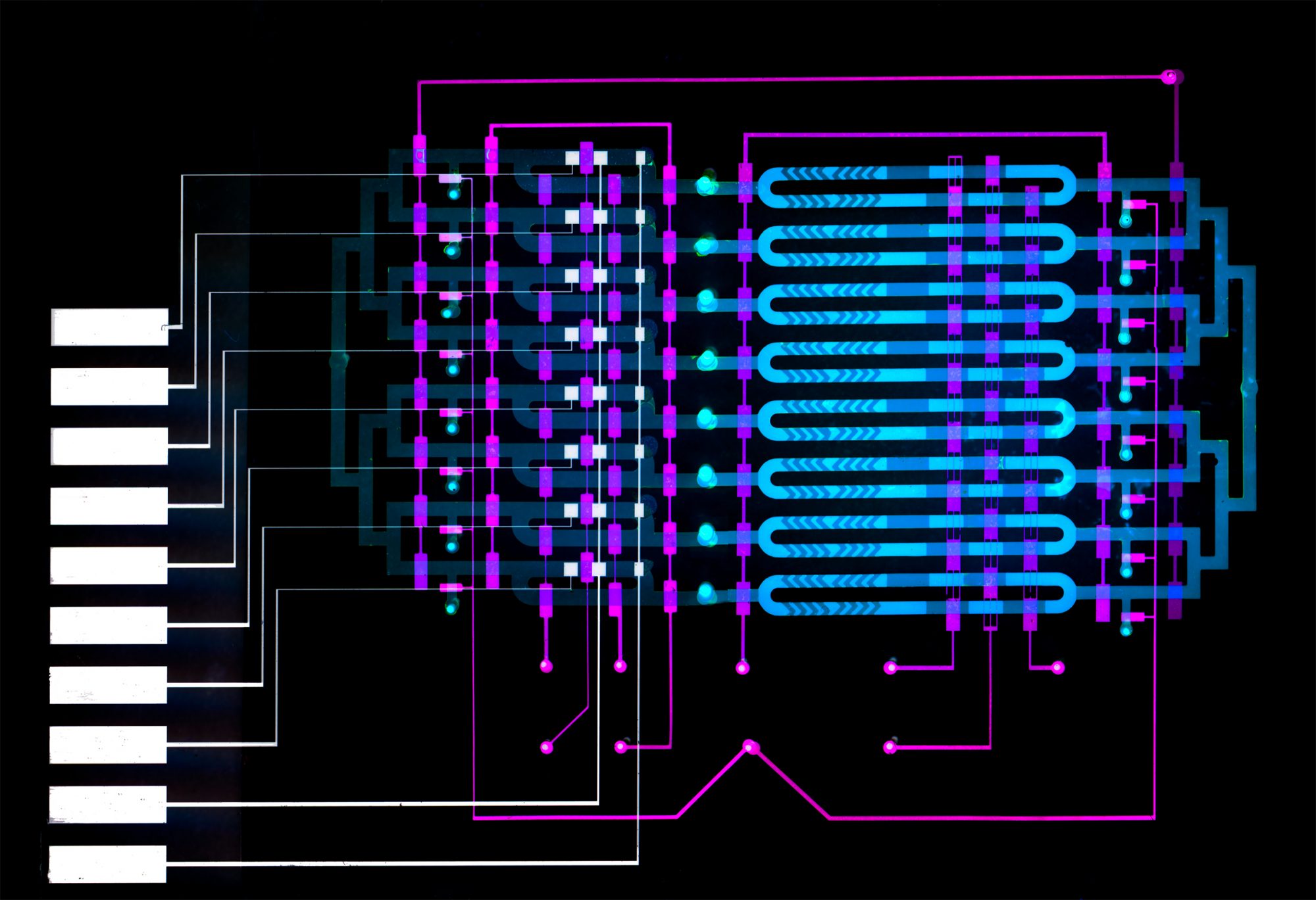
In their work, the researchers wanted to shrink the components of the magnetic bead test, which is often used in the laboratory, on an automated microfluidic device of several square centimeters. This required manipulating beads in micron sized channels and making a device in the Microsystems Technology Laboratory that automates the movement of fluids.
The beads are coated with an antibody that attracts IL-6, as well as a catalyst enzyme called horseradish peroxidase. The beads and the blood sample are injected into the device, entering an "badyte capture area," which is essentially a loop. Along the loop is a peristaltic pump – commonly used to control liquids – with valves automatically controlled by an external circuit. The opening and closing of the valves in specific sequences circulates the blood and the balls to be mixed. After about 10 minutes, the IL-6 proteins bound to the antibodies on the beads.
The automatic reconfiguration of the valves at that time forces the mixture into a smaller loop called the "detection zone", where they remain trapped. A small magnet collects the beads for a brief wash before releasing them around the loop. After about 10 minutes, many beads stuck on an electrode coated with a separate antibody that attracts IL-6. At this point, a solution flows into the loop and wash the loose beads, while those containing the IL-6 protein remain on the electrode.
The solution contains a specific molecule that reacts with the horseradish enzyme to create a compound that responds to electricity. When a voltage is applied to the solution, each remaining bead creates a small current. A common chemistry technique called "amperometry" converts this current into a readable signal. The device counts the signals and calculates the IL-6 concentration.
"For their part, doctors are just loading a blood sample with the help of a pipette. Then, they press a button and 25 minutes later, they know the concentration of IL-6, "says Wu.
The device uses about 5 microliters of blood, which is about a quarter of the finger-prick blood volume and a fraction of the 100 microliters needed to detect protein biomarkers in laboratory badays. The device registers IL-6 concentrations as low as 16 picograms per milliliter, which is lower than the concentrations that signal sepsis, meaning that the device is sensitive enough to allow clinically relevant detection.
A general platform
The current design includes eight distinct microfluidic channels for measuring as many different biomarkers or blood samples in parallel. Different antibodies and enzymes can be used in separate channels to detect different biomarkers, or different antibodies can be used in the same channel to detect multiple biomarkers simultaneously.
The researchers then plan to create a panel of important biomarkers of sepsis that the device to be captured will capture, including interleukin-6, interleukin-8, C-reactive protein, and procalcitonin. But there is really no limit to the number of different biomarkers that the device can measure, regardless of the disease, Wu says. Notably, more than 200 protein biomarkers for various diseases and conditions have been approved by the US Food and Drug Administration. Drug Administration.
"This is a very general platform," says Wu. "If you want to increase the physical footprint of the device, you can increase the size and design more channels to detect as many biomarkers as you want."
The work was funded by Analog Devices, Maxim Integrated and Novartis Biomedical Research Institutes.
From the publisher advisable Articles
Source link