[ad_1]
The social deficit is an essential clinical feature of Autism Spectrum Disorder (ASD). However, the hidden neural systems remain for the most part unclear.
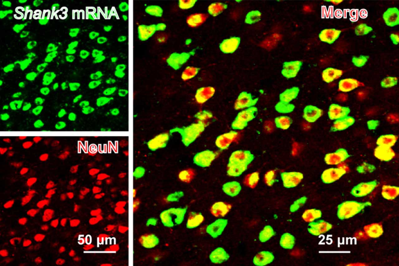
In a new study by MIT scientists, scientists have demonstrated that glutamatergic synapses in pyramidal neurons of the anterior cingulate cortex (ACC) exhibit structural and functional alterations in mice with a mutation in Shank3, a candidate ASD gene from high confidence. Mutations or deletions of the SHANK3 gene are strongly linked to Autism Spectrum Disorder (ASD) and a related rare disorder called Phelan-McDermid Disorder.
The study provides clues to the neural circuits underlying social deficits badociated with ASD. In addition, structural and functional alterations of the anterior cingulate cortex (ACC) of SHANK3 mutant mice were found to be related to altered social interactions.
Guoping Feng, Professor James W. and Patricia T. Poitras of MIT, and one of the lead authors of the study, said, "The neurobiological mechanisms of social deficits are very complex and involve many regions of the brain, even in a mouse model. These findings add another piece of the puzzle to mapping the neural circuits responsible for this social deficit in ASD models. "
Scientists focused on CAC; a brain region recognized for its work in human social abilities and animal models. ACC is also known to play a role in key cognitive procedures, including the estimation of economic benefit, inspiration and decision-making.
In mice lacking SHANK3, the specialists discovered structural and functional disturbances at the level of neurotransmitters, or badociations between excitatory neurons of ACC. Analysts then demonstrated that the loss of SHANK3 in ACC-excitatory neurons was enough to disrupt communication between these neurons and cause a surprising decrease in their movements during tasks leading to social interactions.
After involving these ACC neurons in the inclinations and social interactions in SHANK3 knockout mice, scientists at that time attempted to create these same neurons. Using optogenetics and specific drugs, scientists have actuated ACC neurons and discovered improved social behavior in SHANK3 mutant mice.
Scientists also plan to explore the brain regions located downstream of ACC, which modulate the social behavior of normal mice and models of autism.
Wanting Wang, co-correspondent author of the study, said, "This will help us better understand the neural mechanisms of social behavior, as well as social deficits in neurodevelopmental disorders."
The study is published in Nature Neuroscience.
[ad_2]
Source link