[ad_1]
The Norris Cotton Cancer Center (NCCC) in Dartmouth and Dartmouth-Hitchcock is the first cancer center in the world to install BeamSite Cherenkov imaging cameras in its radiation therapy treatment rooms. The camera system, invented, validated and marketed by entrepreneurs at NCCC and Dartmouth-based biomedical technology company DoseOptics, LLC, captures real-time imaging and video from the beam directly to the patient, allowing the ‘radiation oncology team to visualize the administration of treatment.
Cherenkov imaging turns radiation therapy into a visual process. The Cherenkov effect occurs when beams of photons or electrons interact with tissues, such as skin, producing a small emission of light from the surface. BeamSite cameras can capture images of treatment beam shapes in real time, as well as show intensity levels proportional to radiation dose. This visual data can be used to verify both the accuracy of dose and beam delivery at each daily treatment, which cannot be verified using standard quality assurance measures.
“Cherenkov imaging helps visualize radiation therapy treatment, so the treatment team can see everything and make adjustments when unexpected things happen,” says Brian Pogue, PhD, co-director of the Translational Engineering Research Program. on cancer at NCCC, MacLean Professor of Engineering at Dartmouth Engineering and co-founder of DoseOptics, LLC. A joint engineering and oncology team reviewed the events recorded in their Cherenkov imaging study over the course of a few years, during which they documented incidents when radiotherapy delivery was not ideal and adjustments made to remedy it. Their results, “Initial clinical experience of Cherenkov imaging in external beam radiotherapy identifies possibilities for improving treatment delivery,” were published in The International Journal of Radiation Oncology, Biology, Physics.
There were a total of 64 patients in the study, under the supervision of radiation oncologist and lead author Lesley Jarvis, MD, PhD, member of the Translational Engineering Program in Cancer Research at NCCC and Associate Professor of Medicine at Dartmouth Geisel School of Medicine. The patients were receiving treatment for breast cancer, sarcoma, lymphoma and other cancers. Six patients reported that adjustments would have improved treatment, such as exposure to a dose of stray radiation to the opposite breast, arm or chin during breast cancer treatments. The imaging system was also used to identify when inadvertent dose was not an issue, such as confirmation of no unintentional exposure of the opposite leg during treatment for sarcoma of the extremities.
Radiation therapy is a repetitive procedure administered to patients daily for about 30 days. Placing patients on the treatment table and daily alignment of the beam is a complex process. Beyond postural complications, the therapy team must leave the room when the beam is on, so if anything happens during childbirth, the problem-solving tools are very limited. National statistics show that incorrect delivery incidents can occur at a level of around 1%. In a busy clinic, that could mean one patient per week. “Usually the treatments are very good,” Pogue says. “However, if you can’t see where the beam is, then this is a blind treatment, and the interaction between the patient and the therapy team is just less natural than it is. could not be if the processing was visual. “
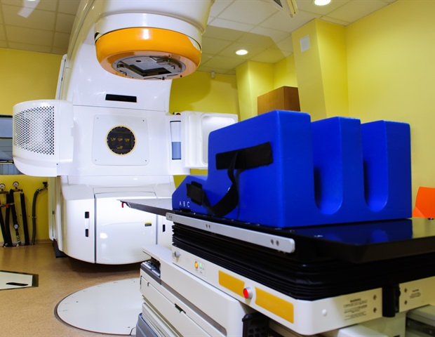
The NCCC is currently the only cancer center in the world to regularly use Cherenkov imaging in all radiation therapy treatments, and was ideally positioned for clinical research teams to test these cameras for the planned study. Cherenkov imaging cameras have been installed in most of Dartmouth-Hitchcock’s linear accelerators, providing an additional level of safety during each patient’s therapy session. “Cherenkov cameras mounted inside radiation therapy treatment rooms allow us to simply see the treatment and provide an intuitive guide to therapists that we would not have had otherwise,” Pogue says. “It’s a great tool for keeping track of what’s happening every day and in every treatment, and for improving the quality of radiotherapy delivery.”
DoseOptics’ technology was developed through research at Dartmouth by the Faculty of Dartmouth, which subsequently licensed the product to the company. Started in Dartmouth-Hitchcock, it is now expanding to other cancer centers. Since DoseOptics, LLC, received FDA clearance to market BeamSite in December 2020, the team has been hoping that all radiation oncology clinics will introduce the technology into their programs. “Clinics should have all the tools at their disposal to make sure that every treatment for every patient is accurate, and to be able to quickly spot problems and resolve them,” Pogue says.
Brian W. Pogue, PhD, is Co-Director of the Translational Engineering Program in Cancer Research at the Norris Cotton Cancer Center in Dartmouth and Dartmouth-Hitchcock, MacLean Professor of Engineering at Dartmouth Engineering, Professor of Surgery at the Geisel School of Medicine of Dartmouth, and President and co-founder of DoseOptics, LLC, which develops camera systems and software for radiotherapy imaging of Cherenkov light for dosimetry. His research interests include optics in medicine, biomedical imaging to guide cancer therapy, molecular guided surgery, dose imaging in radiotherapy, Cherenkov light imaging, image guided spectroscopy. of cancer, photodynamic therapy and modeling of the pathophysiology and contrast of tumors.
Source:
Dartmouth-Hitchcock Medical Center
Journal reference:
Jarvis, LA, et al. (2020) Initial clinical experience with Cherenkov imaging in external beam radiation therapy identifies opportunities to improve treatment delivery. International Journal of Radiation Oncology * Biology * Physics. doi.org/10.1016/j.ijrobp.2020.11.013.
Source link