[ad_1]
A new open source method of bioimprint represents a major breakthrough in the field of regenerative medicine and its success comes from a special ingredient: food coloring.
This week's Science cover presents a spectacular network of entangled hydrogels resembling blood vessels, which is one of many constructs created by a team of bioengineers from Rice University, University Washington, Duke University, Rowan University and Nervous System in Somerville, Mbadachusetts).
The group overcame major obstacles in bioengineering, which freed critical design freedoms in the world of bio-printing, as evidenced by the other achievements presented today in Science. The method was published as an open source resource and allowed for the development of a bioinspired alveolar model, a 3D functional bicuspid venous valve, and a support for grafted liver tissue in a mouse model of chronic liver injury. .
Overcoming one of the biggest hurdles of regenerative medicine
Since the number of people waiting for an organ transplant far exceeds the number of organs available, alternative solutions are needed. Therefore, an important interest lies in the field of regenerative medicine and bioimpression, which could theoretically allow the printing of replacement organs from the patient's cells, thus reducing the risk of organ rejection.
Kelly Stevens, co-author and badistant professor in the Departments of Bioengineering and Pathology at the University of Washington, believes this theoretical solution will become a reality:
"We envision bioprinting to become a major component of medicine in the next two decades."
The inability to provide oxygen and nutrients to all cells of an artificial organ or tissue transplant is one of the greatest challenges in generating functional tissue replacements. Without blood vessels to provide nutrients and eliminate waste, the tissues will not survive very long. As a result, the team sought a method of printing the intricate vascular system that would allow tissues to grow, explained Jordan Miller, co-author and adjunct professor in the Department of Bioengineering at the University. Rice:
"The general idea of our field is to try to understand the structure of human tissues. And I think that one of the things that is really lacking in our field is a part of this multivascular architecture that we have in the body. So, if we hope to build something that looks like the human lung, we do not need a single network of ships, we need at least two because we need lanes. respiratory and blood circulation. "
Food Color Forms Regenerative Medicine
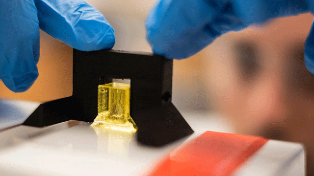
To build the soft vessels, a form of 3D printing called "stereolithography" was used. It relies on light to allow the monomers to bind. This process can be tightly controlled. while the xy resolution is determined by the light path, the resolution z is dictated by additives that absorb excess light and limit the polymerization to the desired layer thickness.
However, it has not been easy to find the right light blocking additives. This method is used in the manufacture of plastic parts, but the additives contain known genotoxic and carcinogenic properties – certainly not suitable for bioproduction! As a result, the team looked for nontoxic light blockers. They tested some promising organic candidates: yellow food coloring, curcumin (based on turmeric) and anthocyanin (based on blueberries), as well as inorganic gold nanoparticles (50 nm) known for their biocompatibility properties and Attenuation of light.
 Daniel Sazer, a bioengineer at Rice University, is developing a lung-scale model of a lung-breathing bag for testing. In experiments, the air is pumped into the bag in a pattern that mimics breathing, while blood flows through a network of surrounding blood vessels to oxygenate human red blood cells. (Image credit: Jeff Fitlow / Rice University)
For these studies, yellow food coloring (tartrazine) was chosen as the winner of the photoabsorbent competition. Miller commented what it meant for the project:
"Thanks to this innovation in the use of these food dyes as light blockers, we are enjoying a new freedom of vascular design. We are able to make many different things with these soft, water-based hydrogels that we could not make before. "
A big step forward for tissue engineering of biomaterials
Thanks to Nervous System's newly found creative freedom and expertise in design, a design studio at the crossroads of science, art and technology, the group has come up with a synthetic vascular system based on transparent hydrogels. In addition, the vessels were incorporated around a functional imitator of lung tissue that was 3D printed using the same technique.
The vessel / lung structure has achieved some significant tests:
- The ships were strong enough to withstand the "breathing" movement of the alveolar models
- The alveolar model provided oxygen to the red blood cells that pbaded through the vessels
- 3D bicuspid valves have been incorporated to respond quickly to changes in flow direction
Miller recalls one of the most exciting moments of the project:
"It was the first time we used the pneumatic system to ventilate the airways. It really looked like a breath … and the complexity of the architecture was so amazing that we immediately saw new things in the blood, as soon as we started doing it. This really transformed our vision of the possible thanks to soft materials containing more than 80% of water. "
Video Credit: Rice University
In anticipation of replacement organs
To better mimic biochemical conditions in vivo, the group showed that endothelial cells injected into the airways could line the vessels and survive. Miller says the group is looking to do similar types of experiments in blood vessel architectures.
"We have shown that human cells can survive in the pre-hydrogel mixture, in the polymerization process and in the functional gel we have made. One of the studies we inserted in this supplement was on mesenchymal stem cells. We were able to show that they were able to survive, grow and spread in 3D inside the frost. They were also able to differentiate into a bone-like cell line beginning to prepare to mineralize this matrix. We really see the potential of this technique not only for modeling living tissue, but also for constructing it and understanding its function in the body. "
To test the relevance of this approach to therapeutic implants, the team printed 3D hydrogel carriers, loaded them with hepatocyte aggregates and implanted them into models of chronic liver injury in mice. After 14 days of transplantation, there were positive signs; there were signs of surviving functional hepatocytes and host blood was present in the explanted tissues.
 Researchers from Rice University and the University of Washington conducted experiments to determine if hepatocytes would function normally if they were incorporated into a bioprinted implant and implanted surgically in the mouse for 14 days. (Image credit: Jordan Miller / Rice University).
Stevens shares his plans for the future: "Here we tested a single function of the liver cells. But liver cells have about 500 functions. In the future, we will test whether our bio-printed tissues can fulfill many more of these functions. We are also working to improve the resolution of our pattern, which is still an order of magnitude greater than the size of most human cells. "
Science and art: a dream wedding
A key element of this work has been the contribution of Nervous System, which, according to Miller, will help transform the field:
"They wrote their own software to build generative architectures, based on algorithms that people observe in nature. And so my suggestion a few years ago was that we could use their algorithms not only to build structures that look like living tissue but could actually be used to make living tissue. "
Jessica Rosenkrantz, co-founder and creative director of Nervous System (Twitter: @nervous_jessica, Instagram: @nervous_jessica) reflects on the occasion:
"As designers whose work inspires complex patterns that we see in nature, it's a dream come true that to be able to work on designing living objects." In our studio, we develop generative software for the design of complex geometries. These types of tools are ideal for tissue engineering where we need to create complex and customized structures such as the vascular system. "

An American dollar coin shown next to a model of respiratory bag mimicking the lungs with airways and blood vessels. Image credit: Brandon Martin / Rice University
"We believe science should be open-source"
This work represents a significant breakthrough in the field of bioengineering and regenerative medicine and opens doors to work on microfluidics, organ-on-chip pathology models and drug discovery.
Miller recognizes the value of open source technology and is pleased to be able to give back to the community that contributed to this project:
"Another interesting part of this work is that all of our equipment used to both 3D print the vessel structures in the gel and perform the ventilation – all this is open source. Later in the day, users will be able to download it legally and freely. I think this will allow other people to really explore this space like never before. "
Reference:
Grigoryan, B., Paulsen, D., Corbett, D., D. Sazer, C., Fortin, A., Zaita, A., Greenfield, P., N., Calafat, J., Gounley, H., Anderson, H. Johansson, F., Randles, A., J. Rosenkrantz, J., Louis-Rosenberg, P., P., K. Stevens, and J. Miller. (2019). Multivascular networks and functional intravascular topologies within biocompatible hydrogels. Science doi: 10.1126 / science.aav9750
[ad_2]
Source link