[ad_1]
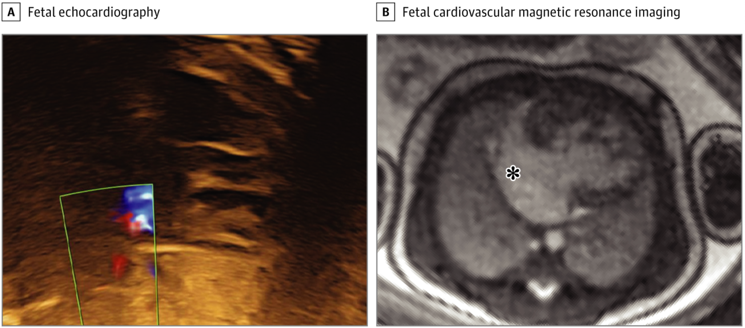
When echocardiography results are unclear, fetal cardiovascular MRI (CMR) can provide valuable information about congenital heart defects, leading to changes in treatment or management decisions in over 80% of cases. , according to a new study.
Not only could this imaging information be used to augment treatment for infants, a team from Lund University in Sweden said, but it could also be useful in providing advice to parents. In fact, they said, 84% of the cases referred for fetal CMR captured clinically valuable data that impacted delivery mode choices, early postnatal care planning, and parental counseling.
“In this study, fetal CMR added clinically useful information to echocardiography in referred cases and was a useful adjunct to fetal echocardiography to assess cardiac function and intracardiac and vascular anatomy in patients. [congenital heart defects]Said the team led by Daniel Salehi, MD, doctoral student in clinical physiology at Lund University.
The team published their results in JAMA network open.
Congenital heart defects (CHD) are rare, occurring in only 1 percent of children. However, they are responsible for about 30% of infant deaths due to abnormalities from birth, and 25% of all cases involve complex abnormalities that require intervention in the first year of life.
Echocardiography usually detects coronary heart disease in utero, but if the acoustic window is inadequate, the results of the scan will not be conclusive. An MR-compatible Doppler ultrasound machine can work around this problem to capture high-resolution fetal CMR images without the need for post-processing, the team said.
Related Content: 4D MRI: A “Massive Leap Forward” in Fetal Cardiac MRI Imaging
To determine the performance of fetal CMR after inconclusive echocardiographic studies – and whether new information guided parenting decisions – Salehi’s team looked at the results of 31 fetuses, with a mean gestational age of 36 weeks, who have been referred for fetal CMR to Skåne University Hospital between January 2017 and June 2020.
In addition to contributing to decision-making and parenting counseling in 84% of cases, the team also determined that fetal RMC was also providing useful information in other cases:
- Intracardiac anatomy and ventricular function were visualized in 87% of fetuses assessed for univentricular versus biventricular outcome in borderline left ventricle, unbalanced inter-atrioventricular communication, and pulmonary atresia with intact ventricular septum.
- Diagnostic information was added in 80% of the aortic arch anatomy cases, including signs of coarctation.
- A delivery planning assistant was provided for 75% of fetuses with hypoplastic left heart syndrome.
- Valuable information for parenting counseling was offered in 68 percent of cases.
“These results suggest that fetal cardiovascular magnetic resonance imaging may add important diagnostic information and affect clinical decision making and parental counseling,” the team said.
But, even with these benefits with fetal CMR, echocardiography still has a role to play, said Bhawna Arya, MD, attending physician and assistant professor of pediatrics at Seattle Children’s Hospital in an accompanying editorial. Fetal echocardiography is useful for identifying coronary heart disease as early as 12 weeks gestation – fetal CMR is relegated to later gestation and larger fetal size, she said.
“Although fetal CMR offers an attractive opportunity for advanced and late gestational imaging, fetal echocardiography remains the gold standard for the early and accurate in utero diagnosis and monitoring of congenital heart disease and other diseases. fetal cardiovascular disease, ”she says.
For more coverage based on knowledge and research from industry experts, subscribe to the electronic diagnostic imaging newsletter here.
Source link