[ad_1]
A team of seven Ghanaian doctors performed the first-ever brain surgery in the country, without cutting the skull.
Technically known as "endovascular spiraling of the brain" (because it is not necessary to cut the skull to perform surgery), the technique pbades a tube (catheter) through the groin (junction zone between the abdomen and thigh on both sides). pubic bone) in the artery containing the cerebral aneurysm, a process known as endovascular coil or spiral embolization.
The surgery, which lasted two hours, was performed with modern equipment and computer software at the Advanced Diagnostic and Cardiology Center at Euracare, a private health facility in Accra.
Dr. Benjamin Dabo Sarkodie, Interventional Radiologist and also Head of Radiology at Euracare, led the team.
Ghana News Titles
For the latest news in Ghana, visit the Graphic Online titles page
Ghana News Page
The other doctors came from the Stroke Unit of Korle Bu University Hospital and Euracare.
Supported by a visiting radiologist, Dr. Itsvan Lazar of Hungary, the team performed a minimally invasive surgery of the brain to stop bleeding, a condition known as "subarachnoid hemorrhage".
This procedure used minimizes surgical cuts to reduce body trauma.
In an interview with The Daily Graphic in Accra nearly three weeks after the operation, Dr. Sarkodie stated that the patient, who was suffering from a broken brain aneurysm, had been discharged a few days earlier and was recovering.
The condition, which is fatal, affects men and women of all ages.

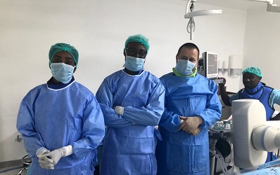
Dr. Benjamin Sarkodie (right) with the support of Dr. Itsvan Lazar (left) and other doctors have undergone successful surgery.
What is a cerebral aneurysm?
Cerebral aneurysm, also known as
This area of the blood vessel uses
Dr. Sarkodie explained that, due to the weakness of the aneurysmal wall, there was a risk of sudden bursting of the aneurysm, a life-threatening situation.
He added that the condition could lead to reduced blood flow to the brain, too much cerebrospinal fluid in the brain, coma, irreversible brain damage and death.
Most cases (about 90%), he said, showed no symptoms because the blood vessels that caused it
Surgery
The team performed an endovascular coil or embolization of the coil, which means that it was not necessary to cut the skull to perform the operation.
Instead, a catheter was advanced from a blood vessel in the groin into the blood vessels in the brain, and
Once the catheter was in place, tiny platinum coils were introduced into the aneurysm.
The tiny platinum soft coils visible on X-rays follow the shape of the aneurysm.
The wound aneurysm becomes coagulated (known as embolization), thus preventing rupture.
sYMPTOMS
According to Dr. Sarkodie, although a cerebral aneurysm may be present without symptoms, the most common initial symptom was a sudden headache caused by subarachnoid hemorrhage (SAH), which is a type of aneurysm. life-threatening stroke caused by bleeding in the space surrounding the brain.
He explained that a serious headache suddenly badociated with HSRA constituted a medical emergency.
The interventional radiologist mentioned the symptoms of an unruptured brain aneurysm, including headaches (rare, if not ruptured), eye pain, vision deficits and deficits in eye mobility.
Causes of cerebral aneurysm
He said that currently the cause of the cerebral aneurysm was not clearly understood, but he insisted that it was badociated with many factors, including smoking, smoking. Hypertension and family history; that is to say genetic.
He added that the ultimate cause of a cerebral aneurysm was an abnormal degenerative change in the wall of an artery and the effects of pressure pulsations of blood pumped forward through the cerebral arteries .
Risk factors
Dr. Sarkodie mentioned that some risk factors badociated with the formation of aneurysms include advanced age, alcohol consumption (especially drinking), atherosclerosis, ie, artery disease characterized by the deposition of fat, such as fibrin on the inner walls.
Others, he said, were smoking, the use of illicit drugs such as cocaine or amphetamine, hypertension (high blood pressure) and trauma or head injuries.
He pointed out, however, that while these risk factors
He added that some people with one or more risk factors had never developed the disease, while others who developed it had no known risk factors.
He stated that "knowing your risk factors for illness can help you choose appropriate actions, including changing behaviors and being the subject of clinical follow-up for the disease."
Diagnostic?
Dr. Sarkodie stated that cerebral aneurysm was often discovered after a rupture or by chance during diagnostic tests, such as
He added that, in addition to complete medical history and physical examination, cerebral aneurysm diagnostic procedures could include digital subtraction angiography (DSA); it is a radioscopic technique used in interventional radiology to clearly visualize the blood vessels in a bone environment or dense soft tissues.
Team
The doctors who practiced the revolutionary surgery were Dr. Benjamin Dabo Sarkodie,
[ad_2]
Source link
