[ad_1]
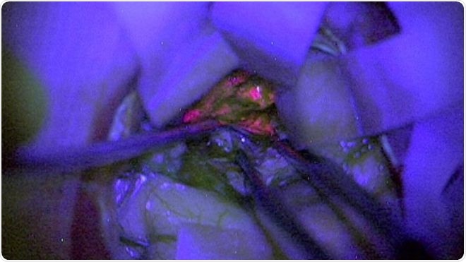
A clinical trial has shown that a special chemical makes brain tumors shiny with pink. This can help neurosurgeons easily operate on them and remove them completely and safely, according to the researchers. The results of this study would be presented at the 2018 NCRI Cancer Conference in Glasgow, 4-6 November 2018.

"Improvement of Intraoperative Diagnosis of High-Grade Gliomas Using a Fluorescence Biomarker – Result of the British NCRI GRIA-BIDD Study, Kathreena Kurian et al., M
For scientists, brain tumors called gliomas are specific: when patients who consume them consume a drink containing 5-aminolevulinic acid or 5-ALA, this one accumulates in The reason behind this is that the tumors do not contain an enzyme necessary for the breakdown of the chemical.This chemical contains a pink color appeared in the tumors that develop most aggressively. differentiate them from healthy brain tissue, he adds, which makes the operation easier and safer.
Patients with glioma are usually treated by surgery to remove r completely the tumor. in these patients. As part of this clinical trial, 99 patients with glioma were included at Royal Liverpool Hospital, Kings College Hospital in London and Adenbrooke's Hospital in Cambridge. They all had high grade gliomas or aggressive growth. Patients received a drink containing 5-ALA just before the operation.
During the operation, surgeons examined the fluorescent tissue of the brain containing the tumor tissue. In 85 patients, they were clearly able to see the tumor tissue as a bright pink. Of these, 81 were in fact high-grade gliomas, confirmed by pathological specimens taken from the brain. One of the glowing tumors has proven to be a low grade tumor. Of the 14 patients in whom the tumors did not shine, seven were low grade and seven were not evaluable for severity.
According to researcher Kathreena Kurian, badociate professor of brain tumor research at the University of Bristol, this type of operational marker was "urgent". She said it would help neurosurgeons find the worst. "The beauty of 5-ALA is that it allows you to see where the high-grade gliomas are in operation," she said.
Principal Investigator Colin Watts, Professor of Neurosurgery and Chair of the Birmingham Brain Cancer Program The University of Birmingham has stated that it is important for neurosurgeons to distinguish cancerous tissue from healthy tissue. This is the first prospective trial demonstrating the benefits of using 5-ALA to improve the diagnostic accuracy of high-grade glioma during surgery. These results show that the marker very well indicates the presence and location of high grade cancer cells. "The advantage of this technique is that it helps to more quickly highlight high-grade disease in a tumor during neurosurgery." This means that a larger part of the tumor can be removed more safely and with fewer complications, which is better for the patient, "he explained.
The team then wants to examine patients with low-grade tumors and badyze how this tool could be used among them. They explain that for low grade tumors, other markers might be helpful. Researchers are also planning clinical trials on this marker in children with glioma. They also plan to evaluate whether this marker can help detect tumors that have returned to the brain after being removed successfully earlier.
Dr. Paul Brennan, of Cancer Research UK, said, "Emphasizing more aggressive tumor cells from real-time treatment could help doctors find the right balance between removing as much of the tumor as possible while preserving surrounding healthy tissue. The fluorescent marker can also reduce the burden of follow-up treatment because cancer cells left behind after surgery require additional radiotherapy or chemotherapy. "
The National Institute of Health and Clinical Excellence also approved the use of 5-ALA. In patients with brain tumors before the operation this year
Source:
https://www.ncri.org.uk/wp-content/uploads/2018/11/Kurian-Glioma-for-online.pdf [19659014] //
[ad_2]
Source link