[ad_1]
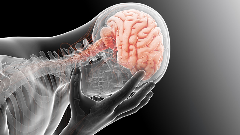
Increasing communication between the amygdala and hippocampus appears to correlate with symptoms of depression and anxiety,
Combining electroencephalography (EEG) recordings and self-reported mood data with machine learning methods, the researchers discovered an "amygdala-hippocampus subnetwork" that correlates with individual variation in mood
"Our findings contribute to a deeper understanding of the neural encoding of mood and anxiety Treatment of mood and anxiety disorders, in particular, innovative treatments using closed-loop deep brain stimulation, "the authors write."
"We found that worsening mood is badociated with increased communication between the amygdala and hippocampus, which have long been linked. co-senior author Vikaas Sohal, MD, PhD, psychiatrist and neuroscientist University of California San Francisco (UCSF), Medscape Medical News.
"This is very intriguing." "But this is very intriguing." But future studies could test this idea, "said Sohal.
The study was published online November 8 in Cell .
Unique Dataset
The researchers took advantage of a unique dataset: intracranial EEG recordings of the limbic system collected over several days in 21 patients
In comparison brainwave activity with the mood diaries, 13 of 21 patients showed fluctuations in electrical activity (or communication) 13-30 cycles / second between the amygdala and hippocampus that correlated with depressed mood. These 13 patients also had significantly higher anxiety than the eight patients in whom this amygdala-hippocampus subnetwork was absent.
The results point to a "specific spatiotemporal neural signature that encodes a large portion (roughly 40-50%) of the Variation in mood over time. This subnetwork is shared over 60% of individuals and is consistently present in individuals with elevated anxiety, "the authors write."
"This research is the first step in letting us look at how the brain (1995) "We do not know whether or not a physician is a patient or a patient who is not a patient."
"If this brain activity" (19659005), "Sohal told Medscape Medical News . between the amygdala and hippocampus) causes causes of confusion, then it could be possible to look at these two structures, "she explained.
" alternative, if this brain activity does not It may be possible to monitor this signal in a patient's mood, but it may be possible to monitor this signal in patients with severe depression. This is the case for the treatment of heart rhythm which is dangerously abnormal, "Sohal suggests.
New Treatment Avenue?
Commenting on the findings for Medscape Medical News , Conor Liston, MD, PhD, neuroscientist and psychiatrist in the Feil Family Brain & Mind Research Institute at Weill Cornell Medicine in New York City, said the research provides a potential new avenue for treatment
just do not understand the neurobiological basis for it in any great detail yet. What's really exciting about this comment? "Said Liston.
" What's really exciting about it? This is the first time it's been introduced to the world, but it's still going strong, but it's still going strong, "he added.
Helen Mayberg, MD The New York City Center for Advanced Therapeutics, IMAH School of Medicine at Mount Sinai in New York City, said this is "really interesting work, capitalizing on the opportunity afforded by chronic recordings in epilepsy patients."
Mayberg said the findings have implications for "depression, anxiety, and post-traumatic stress disorder."
She said her team will be "readin This study is continuing to look at our own chronic undergoing treatment in patients with depression. We can now do long-term recordings from deep brain stimulation leads. This paper provides a clear strategy for the field, "she said.
The research was funded by the Systems-Based Neurotechnology for Emerging Therapies (SUBNETS) program of the Advanced Defense Research Projects Agency (DARPA). [Full text]
For more information on Medscape Twitter
[ad_2]
Source link
