[ad_1]
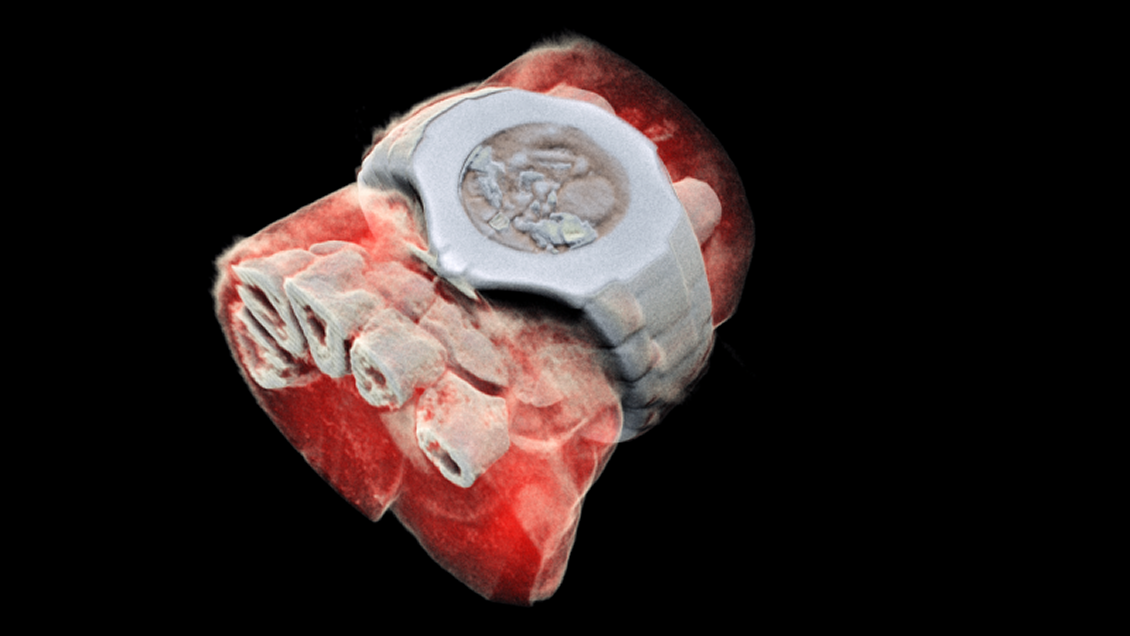
A true revolution for medicine was announced this week by a new technology developed by the European Organization for Nuclear Research (CERN), Medipix3, which has resulted in three-dimensional and color radiographs .
With this technology, the New Zealand company MARS Bioimaging Ltd. scanned, for the first time, a human body with the aid of an advanced color medical scanner that could be used for research, monitoring and treatment
As detailed at CERN , the original concept of the new Medipix technology works like a camera, detecting and counting every particle that strikes pixels when its electronic shutter is open. This allows high resolution, high contrast and very reliable images, which makes it unique for imaging applications in the medical field.
This pixel detection was initially developed to meet the needs of Large Hadron Collider particle tracking and successive generations of chips have demonstrated their great potential apart from high energy physics for more than a year. 20 years old.
Up to now, researchers have been using a small version of the MARS scanner to study cancer, bone and joint health, and vascular diseases that cause heart attacks and accidents
In the coming months, orthopedic patients and rheumatology in New Zealand will be scanned by the revolutionary scanner in a clinical trial that will be the first in the world, the way for a potentially routine use of this new generation equipment.
"It is always satisfying to see our work take advantage of the benefits for patients around the world – real life applications such as this push our efforts to go even further" they said. CERN.
Source link
