[ad_1]

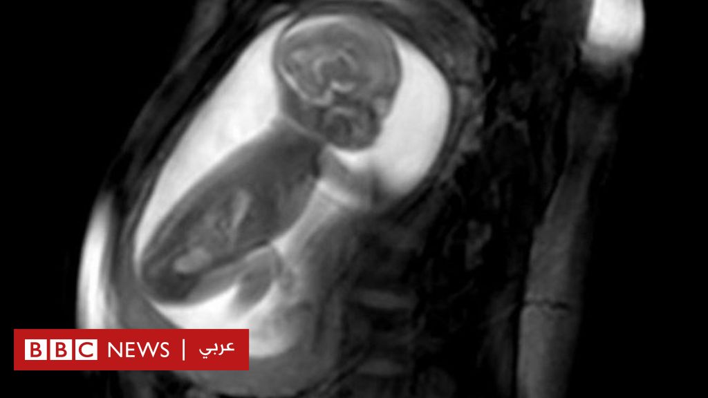
Pregnant women have examined magnetic resonance imaging (MRI) and efficient computers capable of drawing three-dimensional models of the heart of embryos
The researchers managed to take unprecedented pictures of the heart of a fetus beating in the mother's womb, which helped diagnose and treat heart disease immediately after birth.
Pregnant women were examined with magnetic resonance imaging (MRI) and efficient computers capable of drawing three-dimensional models of the heart of small embryos in their mother's womb.
The King & # 39; s College team in London, Guez and St. Thomas confirmed that this new imaging would help improve medical care for fetuses with congenital heart disease.
The researchers say their new achievement can be easily applied in hospitals.
The heart of Violet-Vienna
The Violette-Vienna child was suffering from an unusual health problem in the blood vessels around her heart that was threatening her life while she was in the belly of Kirby's mother, Lea Petit.
Source image
JAMES GALLAGHER
The mother discovered the critical health condition of her fetus during a routine ultrasound examination
The mother discovered the critical health condition of her fetus when she underwent routine ultrasound after 20 weeks of pregnancy.
She then agreed to join the study to allow the researchers to examine in detail the heart of her fetus.
British study: modification of the human embryo genetically "ethically acceptable"
Smoking "night to embryo embryos"
Britain offers free backbone surgeries in her mother's womb
The new technique has shown a narrowing of the main artery from the heart, the aorta (aorta), which would block blood flow into the vessels after birth. Two holes were also found in the heart of the child, which is found in the uterus.
"It was very scary, it was a real shock," Kirby Leah told the BBC.
A BBC journalist holds his heart printed in a 3D printer
But allowed doctors to plan a way to save the life of his child Violet Vienna, after his arrival in the world.
"They did not let me keep them, and they had to put them on the devices immediately to keep the aorta open," Kirby said.
The girl had a heart operation one week after birth. Despite a difficult start, she survived and is now 11 months old.
"It's now only thanks to these researchers and this technology," she said.
"What they do is amazing, it's a thing that saves lives."
How technology works
Researchers take a series of two-dimensional images of the heart from different angles using the MRI device.
But the heart of the fetus is too small, it beats at an incredible speed, and the fetus moves in the womb, so that the image of the heart does not seem quite clear.
But this smart gadget really made the difference.
Advanced computer programs can combine images, adjust them by heartbeat and create an unprecedented three-dimensional image of the whole heart.
It then provides physicians with a clear view of cardiac dysfunction.
Good for children
Congenital anomalies are at the origin of eight out of 1000 children with heart disease in Britain.
Reza Radawi, consultant in pediatric cardiology, wanted to improve the diagnosis of congenital malformations after the birth of a girl infected with a
They can be caused by infection and certain drugs, and can be genetically transmitted through the family.
Professor Rida Radawi, pediatric cardiology consultant, sought to improve the diagnosis of birth defects after the birth of a girl with one of these malformations.
"We thought it would die and it was a strong incentive … we must be able to solve the problem and he is still in the womb," he said.
He describes the three-dimensional images of the heart as "beautiful" and says that they allow doctors to clearly see the problem and to improve care.
"We can reach the stage of greatest certainty (about the problem), plan in advance the necessary treatment and identify the process we need," he told the BBC.
"It really helps parents get the support they need to see what's going on."
"It also really helps children because they have the right surgery on time and therefore the best results."
Will it be used?
The study, published in the Lancet, showed that three-dimensional imaging was a success for 85 pregnant women, but tests have now been performed on more than 200 patients.
Source image
Getty Images
This technique will be easy to apply if the hospital already has an MRI and a computer with a specific graphics card.
"We hope that this approach will become a certified practice for the EEG team and that the team will diagnose antenatal heart disease every year in 400 children," said Dr. David Lloyd, researcher in clinical research at King & # 39; London College.
"This will also improve the medical care of more than 150 children each year, born at St. Thomas Hospital and with known congenital heart disease."
He explains that the technique will be easy to implement if the hospital already has a magnetic resonance imaging device and that the new device it will need is a computer equipped with. an appropriate graphics card.
Future radiology for children?
This research is part of the iFind project to increase the ability to diagnose and diagnose more health problems during standard pregnancy screening.
If these diseases are diagnosed only after birth, you risk losing a lot of time trying to make a correct diagnosis, which increases the risks for the child after birth.
Another method is to simultaneously use four ultrasonic sensors to obtain a more detailed image.
Source link