
[ad_1]
Scientists have found no trace of SARS-CoV-2 in the brains of infected people. However, they observed damage to blood vessels caused by the body’s inflammatory response in the postmortem brain of patients who tested positive for COVID-19, suggesting that the virus could indirectly attack the organ.
Scientific advances made by researchers this year have helped immensely in learning more about the novel coronavirus which first appeared in Wuhan, China.
Initially, COVID-19 was characterized by fever, sore throat, cough and dyspnea, all manifestations of respiratory illness.
However, a spectrum of other clinical manifestations including headache, abdominal pain, diarrhea, and loss of taste and smell have been reported. Researchers also now have evidence that the new coronavirus damages the heart, creates blood clots and causes gastrointestinal problems.
Although the pandemic has disproportionately affected the elderly and those with pre-existing health conditions, young adults are not immune.
Earlier this year, reports showed that young adults who contracted the novel coronavirus had more neurological symptoms, including mental confusion, headaches, dizziness, uncoordinated muscle movements, seizures and increased blood pressure. risk of stroke.
These are just a few examples of everything we have learned and what we still need to learn in order to fully understand the SARS-CoV-2 virus. Now, new research suggests that the virus can also cause brain vascular damage.
The research authors published their findings as a correspondence article in the New England Journal of Medicine.
The study was conducted by researchers at the National Institute of Neurological Disorders and Stroke (NINDS) in Bethesda, MD, and other institutions across the United States.
They examined post-mortem brain tissue samples from 16 patients in New York City and three patients in Iowa City who died between March and July 2020 and tested positive for COVID-19 before or after death.
The patients ranged from 5 to 73 years old and their medical histories frequently showed pre-existing conditions, including obesity, heart disease or high blood pressure, and diabetes.
Before death, treatment was mainly focused on respiratory infections and only two patients presented with agitated delirium.
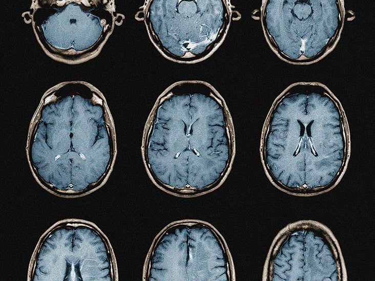
The researchers also used MRI images to detect any abnormalities in the brain tissue. This included the olfactory bulb, an area of the brain involved in smelling, as loss of smell is known to be one of the first symptoms of COVID-19.
Another region of the brain they examined was the brainstem, which is crucial for human survival. It regulates sleep and eating habits and controls heart rate and respiratory rate.
To assess relevant brain tissue, the researchers used a staining method called immunohistochemistry, which allows visualization of proteins in cells and tissues.
Of the 19 brain tissue samples, 13 were imaged and 10 showed brain abnormalities. Further analysis showed damage to the blood vessels.
In nine patients, the presence of lesions suggested that they were lesions of the leaking cerebral vessels. There were also signs of a blood protein called fibrinogen leaking into the brain. The authors suggest that this is evidence of inflammation resulting from an overactive infection-fighting immune system.
In 10 patients, MRI images showed hypointensities corresponding to congested blood vessels and an accumulation of fibrinogen around the area.
“Our results suggest that this may be caused by the body’s inflammatory response to the virus,” says Dr. Avindra Nath, clinical director of NINDS at the National Institutes of Health (NIH) and lead author of the study.
Interestingly, the SARS-CoV-2 virus was not found in any of the brain tissue of the patients. However, the authors write that there is no way to know if the virus was present at any given time:
“It is possible that the virus was cleared at the time of death or that the number of viral copies was below the level of detection by our tests.”
Upon closer examination of the areas of injury, the researchers found that immune cells, such as T cells, were present around the brain, confirming an inflammatory response in the brain.
“We were completely surprised. We originally expected to see damage from a lack of oxygen, ”says Dr. Nath. “Instead, we saw multifocal areas of damage typically associated with stroke and neuro-inflammatory disease.”
With the new coronavirus not having been detected in brain tissue of deceased patients, authors say it is too early to tell if there is a link between the neurological effects associated with COVID-19 and the damage to blood vessels seen In this study.
“In the future, we plan to study how COVID-19 harms blood vessels in the brain and whether it produces some of the short and long term symptoms that we see in patients,” says Dr Nath.
As scientists continue to study the full effects of SARS-CoV-2 infection, it is important to slow the spread of the virus.
The public can help minimize the risk of transmission by wearing a face mask, practicing social distancing, and staying at home as much as possible.
Adherence to these guidelines is essential as immunization efforts continue around the world.
For live updates on the latest developments regarding the novel coronavirus and COVID-19, click here.
[ad_2]
Source link