[ad_1]
Researchers at the University of Cambridge have used organo-culture techniques to create a cellular model of the early stages of the placenta. We hope this will deepen our understanding of the early stages of pregnancy and the etiology of various reproductive disorders.

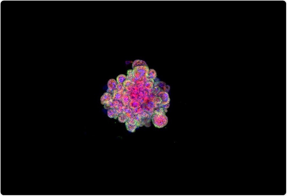
It is a confocal image of a colored organoid trophoblast for cytokeratin 7, F-actin and Dapi. (Credit: Trophoblast Research Center).
The placenta is an organ that develops in early pregnancy. It attaches to the wall of the uterus and the umbilical cord follows from it.
All the oxygen and nutrients that the fetus needs to grow are provided by the placenta and the waste is eliminated by the same route. If the placenta does not develop properly, a series of complications can occur, including stillborn offspring.
The placenta is absolutely essential to support the baby when it grows inside the mother … When it does not work properly, it can result in serious problems, pre-eclampsia to miscarriage, with immediate consequences for the mother and the child. . But our knowledge of this important organ is very limited because of the lack of good experimental models. "
Dr. Margherita Turco, first author
Unsuccessful pregnancies generally result from the fact that the embryo is unable to implant properly and forms a placental attachment.
Although it is known that placental failure is a common cause of infertility, the underlying causes of this failure remain elusive. This is largely a consequence of difficulties in studying the early stages of pregnancy.
Since they can not be studied, we understand very little about what normally happens at the very beginning of our development and how it can go wrong. Unfortunately, animal models are of no use because the processes of placental development and implantation are too different from those of humans.
The University of Cambridge is a world leader in organoids research and has created a range of organ models including 'mini-brains', 'mini-livers' and 'mini' -poumons ". They have now used this technique to generate "mini placentas", which are functional models of the early placenta.
The team developed the "mini-placenta" organoids from villous cells harvested from placental tissues. These trophoblast organoids are very similar to the normal placentas of the first trimester and secrete the same essential proteins and hormones. In addition, they are genetically stable and able to survive in the long term.
Professor Graham Burton, co-author
These "mini-placentas" build on decades of research and we believe that they will transform work in this area. They will play an important role in helping us investigate early pregnancy events that have profound implications for the health of the mother and her offspring throughout life.
Professor Graham Burton, co-author: Organo-placental organelles can help to better understand the relationships between the placenta, the uterus and the fetus and provide a way to check the safety of drugs used in early pregnancy.
The team has already created a miniature functional model of the uterine lining and reported how cells at the junction of the uterus and placenta talk to each other to alter the immune response and allow pregnancy.
Now that we have developed organoid models on both sides of the interface – mother tissue and placenta tissue – we can begin to look at how these two sides speak to each other. "
Professor Ashley Moffett, co-author
sources:
University of Cambridge. Press release of November 28, 2018.
Turco MY et al. Trophoblast organoids as a model of interaction between mother and fetus during human placentation. Nature, Nov 28, 2018
[ad_2]
Source link
