[ad_1]
(Nanowerk NewsPneumonia, a respiratory disease that kills about 50,000 people in the United States each year, can be caused by many microbes, including bacteria and viruses. The rapid detection of pneumonia is essential for effective treatment, especially in cases of hospitalization often more serious. However, current diagnostic methods often take several days to obtain definitive results, which prevents doctors from prescribing the appropriate treatment.
MIT researchers have developed a nanoparticle-based technology that could be used to improve the speed of diagnosis. This type of sensor could also be used to test whether antibiotic therapy has successfully treated the infection, says Sangeeta Bhatia, professor of health sciences and technology engineering and electrical and computer engineering John and Dorothy Wilson, as well as as the lead author of the study.

"If the patient's symptoms go away, then you assume that the medication is acting. But if the patient's symptoms do not go away, then you will want to see if the bacteria is still growing. We were trying to solve this problem, "says Bhatia, who is also a member of the Koch Institute for Cancer Research and MIT's Institute for Medical Engineering and Science.
Graduate student Colin Buss and recent doctoral recipient, Jaideep Dudani, are the lead authors of the article, published online November 29 in the newspaper EBioMedicine ("Protease activity sensors classify non-invasively bacterial infections and antibiotic responses"). Reid Akana, a senior from MIT, and Heather Fleming, research director for Bhatia's lab, are also the authors of the paper.
Sensors in the body
Several years ago, Bhatia and his colleagues developed a diagnostic approach that amplifies the signal of biomarkers already present in the body, particularly enzymes called proteases, which cut out other proteins. The human genome encodes more than 500 different proteases, each targeting different proteins.
The team has developed peptide-coated nanoparticles (short proteins) that can be cut by certain proteases, such as those expressed by cancer cells. When these particles are injected into the body, they accumulate in the tumors, if any, and the proteases hatch the peptides of the nanoparticles. These peptides are eliminated as waste and can be detected by a simple urine test.
"We are working on the idea that measuring enzyme activity could be a new way of looking inside the body," says Bhatia.
In recent studies, she showed that this approach could be used to detect different types of cancers, including very small ovarian tumors, which could allow earlier diagnosis of ovarian cancer.
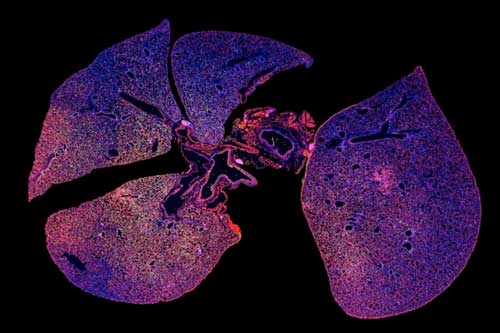
For their new study, researchers wanted to explore the possibility of diagnosing an infection by detecting proteases produced by microbes. They started with a species of bacteria called Pseudomonas aeruginosa, which can cause pneumonia and is a particularly common cause of hospital-acquired cases. Pseudomonas expresses a protease called LasA. The researchers therefore designed nanoparticles containing peptides that could be cleaved by LasA.
Researchers have also developed a second nanoparticle-based sensor capable of monitoring the immune response of the host to infection. These nanoparticles are coated with peptides that are cleaved by a type of protease called elastase, which is produced by immune cells called neutrophils.
In some patients with pneumonia, even if an antibiotic removes the bacteria at the origin of the infection, a chest X-ray can still show inflammation because neutrophils are still active. The joint use of these two sensors could reveal if an antibiotic has eliminated the infection, in cases where a chest X-ray still showed inflammation after treatment.
"Sensors can help you distinguish between infection and inflammation, inflammation and no infection," says Bhatia. "What we showed in the document, is that when you treat with the right antibiotic, the infection decreases, but the inflammation persists."
The researchers also showed that if they treated mice with an ineffective antibiotic, the levels of bacteria and inflammation remained high. This type of test could help determine if an antibiotic is effective in cases where the patient's symptoms have not improved in a few days.
Diagnose many infections
For this study, the researchers delivered the nanoparticles intravenously, but they are now working on a powdered version that can be inhaled.
Bhatia plans to use this approach to determine if a patient has bacterial or viral pneumonia, which would help doctors decide whether the patient should receive antibiotics or not. The definitive test, which consists of culturing a bacterial culture from mucus cough, takes several days. Doctors base their decisions on patient symptoms and radiography, which may not always be accurate.
To create a more complete diagnosis, Bhatia's lab is currently working on adding peptides that can interact with proteases from other types of bacteria that cause pneumonia, as well as proteases produced by the host's immune system. response to a viral or bacterial infection. Researchers are also working on sensors that could easily distinguish between active and dormant forms of TB.
Bhatia and others have started a company called Glympse Bio, which has licensed the use of protease detection technology and is currently developing protease sensors that can be used at home. man. Next year, they plan to start a Phase I clinical trial on a sensor that can detect liver fibrosis, an accumulation of scar tissue that can lead to cirrhosis.
Source link
