[ad_1]
Tech2 News Staff
Nov 24, 2018 4:49 PM IST
A little over ten years ago, two researchers from the University of California at Davis had had the idea of a machine capable of scanning the entire body up and down in one take. The scan would produce a 3D image that could help doctors and medical experts diagnose disorders and develop new treatments faster and better.
This fantasy scanner is now a reality and the first images he produced are much more real than what the researchers expected.
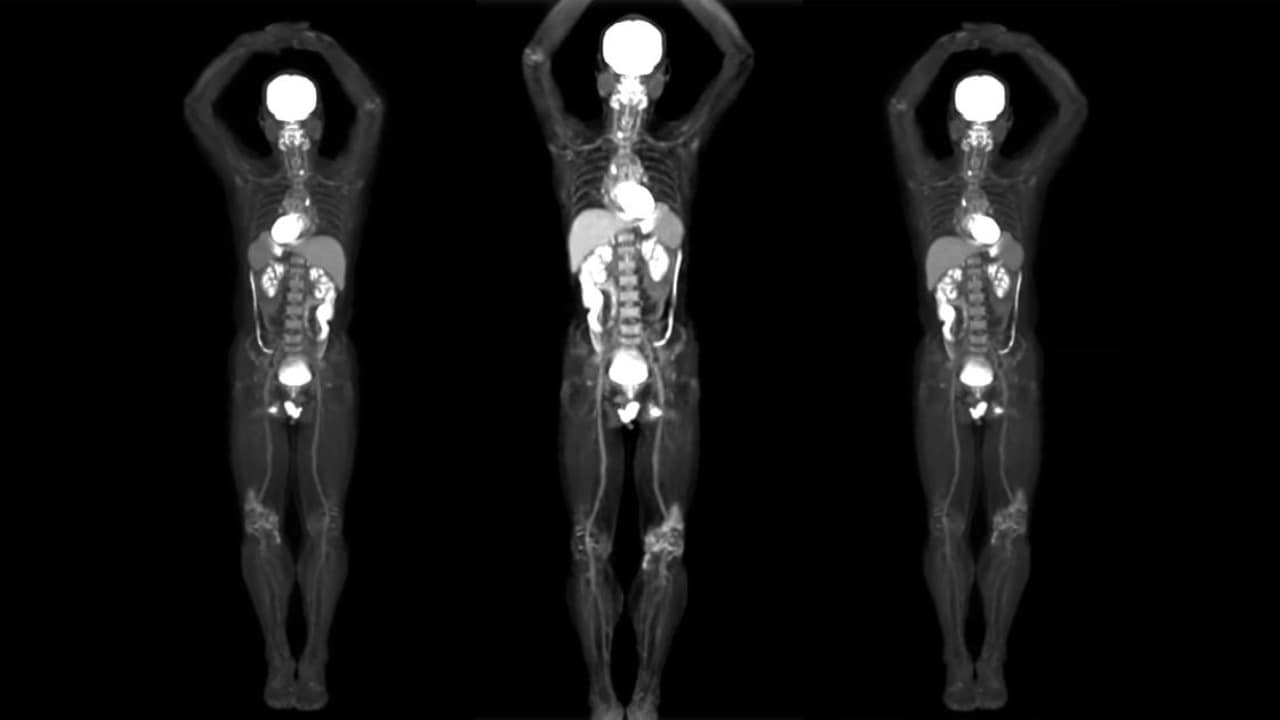
EXPLORER is a scanner that combines two imaging techniques used today – positron emission tomography (PET) and X-ray computed tomography (CT) – to simultaneously examine all organs and tissues from the body.
The biggest advantage of such a scan would be the ability to image multiple areas of the body in one sitting. But that's not all the EXPLORER can do – it's also very, really fast.
While the average scan can take anywhere from 10 to 20 minutes to produce an image, this device can perform a full body scan in just 30 seconds. The researchers also claim that it is much safer and requires a much lower radiation dose than a PET-scan alone.
In addition, EXPLORER can also produce moving body films that can track blood vessels or even drugs that pass through the body.
"Even though I had imagined what the images would look like for years, nothing prepared me for the incredible detail that we could see during this first scan," said Simon Cherry, one engineers from the device. told university press.
"There is no other device capable of obtaining data of this type in humans, so it is really a novelty," added Ramsey Badawi, second searcher.
The duo does not believe that EXPLORER will be used in hospitals and medical centers around the world.
[ad_2]
Source link
