[ad_1]
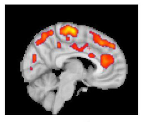
According to the researchers, this combined IR / PET image highlights areas of the brain in which patients with fibromyalgia had increased glial activation, compared to unaffected control volunteers. (Photo courtesy of Marco Loggia, PhD, Martinos Biomedical Imaging Center, Massachusetts General Hospital)
Researchers at Massachusetts General Hospital and Karolinska Institutet in Sweden have documented the widespread inflammation that occurs in the brains of fibromyalgia patients.
Their study is published in the journal Brain, behavior and immunity.
"We do not have good treatment options for fibromyalgia, the identification of a potential treatment goal could lead to the development of innovative and more effective therapies," says Marco Loggia, PhD, of Martinos Center for Biomedical Imaging based at MGH. lead author of the report, in a press release from Massachusetts General Hospital.
"And finding objective neurochemical changes in the brains of patients with fibromyalgia should help reduce the persistent stigma that many patients face, often being told that their symptoms are imaginary and that there is nothing really serious about them. with them."
Fibromyalgia is characterized by symptoms such as generalized chronic pain, sleep problems, fatigue, and problems with thinking and memory.
Earlier research by the Karolinska group led by Eva Kosek, MD, Ph.D., lead co-author of this study, has suggested a potential role for neuroinflammation in this condition – including high levels of inflammatory protein in the fluid cerebrospinal. neuroinflammation visualized directly in fibromyalgia patients.
A study conducted in 2015 by the Loggia team used MRI / PET combined scanning to document neuroinflammation – particularly glial activation – in the brains of patients with chronic back pain. In his study of 20 fibromyalgia patients and 14 control volunteers, his team used the same PET radiopharmaceutical, which binds to the overexpressing translocative protein (TSPO) by activated glial cells. explains the statement.
At the same time, the Kosek team in Karolinska recruited a group of 11 patients and an equal number of participants in a similar study with the TSPO TEP tracer. Since this radiopharmaceutical binds to two types of glial cells – microglia and astrocytes – it also analyzed 11 patients, six of whom had TSPO imaging and five others, and another 11 with PET tracers. microglia. In both centers, participants with fibromyalgia completed questionnaires to assess their symptoms. When the MGH team became aware of the similar survey conducted by the Karolinska group, the teams decided to group their data into one study.
Results from both centers showed that glial activation in several brain regions of fibromyalgia patients was significantly greater than in control participants. Compared to the MGH team's chronic back pain study, TSPO elevations were more prevalent in the brain, which, according to Loggia, corresponds to the more complex symptoms of fibromyalgia.
TSPO levels in a structure called the Cingular Gyrus – an area associated with emotional treatment where neuroinflammation was reported in patients with chronic fatigue syndrome – corresponded to fatigue levels reported by patients. The Karolinska team studies with the astrocyte binding tracer revealed little difference between patients and controls, suggesting that microglia were primarily responsible for increased neuroinflammation in patients with fibromyalgia.
"The activation of glial cells observed in our studies releases inflammatory mediators believed to sensitize pain pathways and contribute to symptoms such as fatigue," says Loggia, an assistant professor of radiology at Harvard Medical School. "The ability to join forces with our colleagues in Karolinska was fantastic because combining our data and achieving similar results at both sites enhances the reliability of our results."
[Source(s): Massachusetts General Hospital, EurekAlert]Source link
