[ad_1]
Researchers at the University of Texas at Austin have demonstrated a new approach to protein sequencing much more sensitive than the current technique..
The new technique makes it possible to identify individual proteins rather than asking for the simultaneous determination of millions of them.
This development could have major implications for biomedical research, facilitating the detection of new biomarkers of disease and improving understanding of healthy tissue function.
This breakthrough is the result of work begun six years ago by Dr. Edward Marcotte and his colleagues, who wanted to adapt the methods used for next-generation sequencing (NGS) to protein sequencing.
In the same way that NGS allowed rapid and accurate sequencing of the entire genome, the technology developed by the team provides fast and detailed information on tens of thousands of proteins involved in health and disease .
We have essentially created DNA sequencing technology to study proteins.
Dr. Edward Marcotte, Principal Investigator
In many diseases such as cancer, diabetes and Alzheimer's disease, cells produce proteins that serve as fingerprints or as unique biomarkers for the disease.
Improving the detection of these biomarkers would help researchers better understand diseases and enable faster and more accurate diagnosis.
The current standard for protein sequencing – mass spectrometry – is often not sensitive and requires the presence of about one million copies of a protein to be detected.
The technique is also a "low flow", detecting only a few thousand types of proteins in a single sample.
As reported in the newspaper Nature Biotechnologythe new method, called single-molecule fluorosequencing, allows millions of individual proteins to be sequenced simultaneously in a single sample.
Marcotte hopes that this number can be increased to billions.
The higher throughput and sensitivity of the new technique compared to the current standard should improve the detection of disease biomarkers and enable scientists to study diseases such as cancer in a whole new way.
Examining a tumor at a cell, for example, could allow researchers to see how a tumor evolves in the form of a small collection of identical cells into a large mass of genetically diverse cells with different properties. . Such knowledge could pave the way for new approaches to targeting cancer.
 Credit: University of Texas at Austin
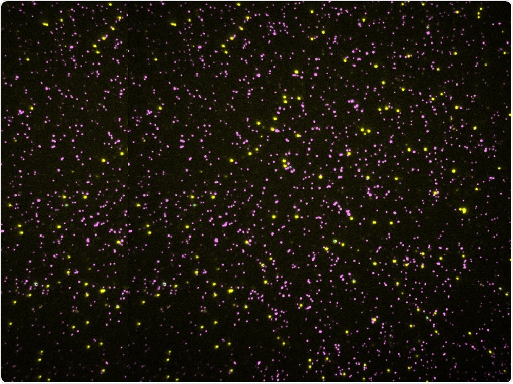
The dots on this image are not stars, but millions of proteins, seen under the microscope. Each protein rises like a blade of grass whose only extremities are visible when we look from above.
Proteins are amino acid chains, so every point is just the amino acid of the tip. Among the 20 types of amino acids that make up proteins, one type is labeled yellow, another is pink, the other 18 are not labeled, they are black.
By removing an amino acid from each protein, taking a new image and then repeating several times, researchers can record an amino acid sequence for each protein in a sample of millions of people simultaneously.
Source:
This report is based on a press release from the University of Texas at Austin.
[ad_2]
Source link