[ad_1]
CHAMPAIGN, Ill., Nov. 6, 2018 – A new molecular probe, based on fluorescence in situ hybridization (FISH) technique, uses compact quantum dots instead of fluorescent dyes to illuminate molecules and diseased cells . Developed by a team from the University of Illinois at Urbana-Champaign and the Mayo Clinic, QDs provide stability and accuracy superior to those of dyes.

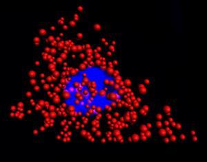
Quantum dots illuminate the locations of individual mRNA as red dots in the cytoplasm of a single HeLa cell. The blue region is the nucleus. This work is the fruit of a collaboration between researchers from the Illinois Bioengineering and the Mayo Clinic. Courtesy of the University of Illinois, Department of Bioengineering of Urbana-Champaign.
Signals of organic dye labels used in FISH deteriorate rapidly during photoexcitation, especially when scanning in 3D and under a high photon flux required for super-resolution. In addition, when dyes are used, the technique is limited to the simultaneous analysis of about three RNA targets at a time.
Until now, size thresholds limited the use of QDs for labeling mRNAs in cells. To overcome this problem, the researchers identified the optimal size that QDs should have to be able to work effectively with the FISH protocol. This discovery led them to develop a FISH based on the QD that corresponds to the labeling specifications currently obtained with organic dyes. The team created QDs composed of an alloy of zinc, selenium, cadmium and mercury and coated with polymers.
"The core of the point dictates the emission wavelength and the hull, the amount of light emitted," said Professor Andrew Smith.
Unlike conventional QDs, team QDs can emit colors regardless of the size of the particle. The QDs measure 7 nm, making them comparable in size to the dyes used in conventional FISH probes and small enough to fit on a probe capable of maneuvering between proteins and DNA in a cell.
In experiments with HeLa cells and prostate cancer cells, the researchers found that the number of dye-based FISH cells decreased rapidly in minutes. The FISH method based on QD provided a long-term luminescence allowing the counting of RNA for more than 10 minutes, allowing the acquisition of 3D cell imaging.
"This is important because cell and tissue images are acquired sequentially, so that later slices labeled with dyes are depleted before they can be replicated," Smith said.
The researchers believe that QD-FISH could be particularly beneficial for high-resolution gene expression studies on 3D biological samples for which quantification and multiplexing are major challenges.
"By replacing dyes with quantum dots, there is no stability problem and we can count many RNAs with higher fidelity than before," Smith said. "In addition, we discovered a fundamental limit to the size of a molecular marker in cells, revealing new design rules for analysis in cells."
This research was part of the Mayo Illinois Alliance in which Illinois engineers work directly with clinicians and biologists from Mayo Clinic to solve outstanding medical problems. The Mayo Clinic's biomarker discovery group is employing to develop FISH-based diagnoses for tumor biopsies to improve the accuracy of cancer diagnosis, select personalized treatments, and improve predictions.
The search was published in Nature Communications (Https://doi.org/10.1038/s41467-018-06740-x).
Source link
