
[ad_1]
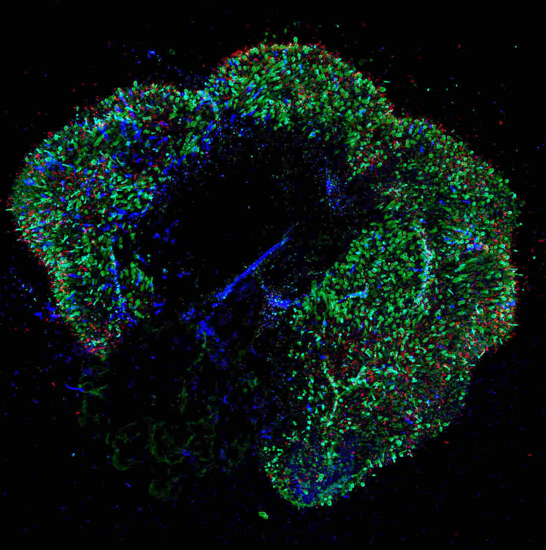
A retina aged 291 days contains photoreceptors in sticks that appear in red. The cells of the blue cone are visible in blue. The red and green cone cells are green.
Johns Hopkins University
hide the legend
activate the legend
Johns Hopkins University

A retina aged 291 days contains photoreceptors in sticks that appear in red. The cells of the blue cone are visible in blue. The red and green cone cells are green.
Johns Hopkins University
To see the red of a sunset or the green of spring leaves, the development of human eyes must get the right hormone at the right time.
It's the discovery of a team of scientists who have studied the development of color vision using hundreds of human-grown retinas in the lab.
The discovery, published Thursday in the newspaper Science, could help accelerate current efforts to treat color blindness. This could also lead to new treatments for diseases such as macular degeneration, the leading cause of vision loss.
"It is important that we understand how nature controls the development of the retina to better understand why disease problems occur and how to treat them," says Steven Becker, a scientist at the National Eye Institute. The recently published results are a step in that direction, says Becker, who has no connection with research.
The development of color vision in people has been difficult to study because it usually occurs in the womb – and out of sight. But two scientists at Johns Hopkins University thought they could find answers using retinas grown in the laboratory.
These "retinal organoids" have been around for a few years, but they are difficult and tedious to develop, says Kiara Eldred, a graduate student at Hopkins, who is the first author of the journal.
Transforming a batch of immature retinal cells into a functional organoid takes up to one year.
"During the first week of their lives, I take care of them every day," says Eldred. After a few weeks, she says, the cells become "a little more independent".
And with a little luck, these groups of immature cells turn into a 3D structure that "looks like a developing retina you would see in a baby," said Bob Johnston, Eldred's boss and assistant professor in the department. of biology.
Johnston's lab had studied vision in flies. But he and Eldred saw a chance to try something much more ambitious.
"We discussed this crazy idea of whether we could use these human retinal organoids to study how we get the different color-sensing cells in our eyes," Johnston said.
The use of human cells was essential because you can not study how humans perceive the colors of a fly or even a mouse.
"The mice do not smell red," says Johnston. "They do not have cells that detect red, so we need to study that in human tissues to get a sense of how it works."
Johnston and Eldred knew that in a human fetus, cells that detect blue light appear first. Then come cells that respond to red and green lights.
And animal research has suggested that the thyroid hormone was involved in the development of these color-detecting cells, called cones. Johnston had Eldred perform an experiment with immature retinal cells.
"I added thyroid hormones to the dish during their development and we had more red-green cones developing in these organoids," she says. "We were really excited because we were on something."
It would take years and many other experiments to confirm that the thyroid hormone was triggering the onset of color vision. And the team still did not understand why some cones became even more specialized by detecting only red or green.
But Johnston says his lab is now gearing up for two new goals.
"One is to restore color vision in color-blind people," he says. What his laboratory is learning, he says, could help accelerate the efforts already made to use gene therapy in the treatment of color blindness.

The second goal of the lab is to use retinal organoids to better understand diseases such as glaucoma and macular degeneration, one of the leading causes of vision loss.
Macular degeneration affects the macula, an area of the retina that provides high resolution vision. The condition has been difficult to study, however, because the mice do not have maculae.
Johnston hopes to learn more by having his lab create macular organoids.
Scientists are increasingly optimistic about the emergence of new efforts to treat retinal diseases, Becker said. At first, researchers had doubts about the fact that a retina grown in a dish could mimic reality.
But studies like this one on color vision, he says, "show that the similarity is quite high."
To encourage scientists to develop more retinal organoids, the National Eye Institute is sponsoring a $ 1 million science competition
Source link
