
[ad_1]
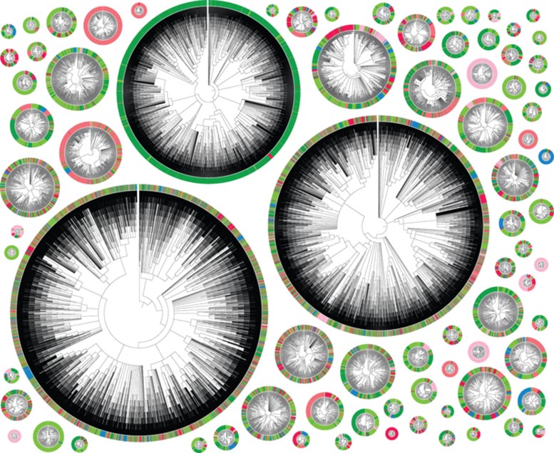
Phylogenetic trees, starting with an individual cancer cell. Each color represents a different location in the body. A very colorful tree shows a highly metastatic phenotype, where the descendants of a cell have jumped between different tissues several times. A tree that is predominantly one color represents a less metastatic cell. Credit: Jeffrey Quinn / Whitehead Institute
Using CRISPR technology, researchers track the lineage of individual cancer cells as they proliferate and metastasize in real time.
When cancer is confined to one place in the body, doctors can often treat it with surgery or other therapies. Much of the mortality associated with cancer, however, is due to its tendency to metastasize, sending seeds of itself that can take root throughout the body. The exact moment of metastasis is fleeting, lost in the millions of divisions that take place in a tumor. “These events are generally impossible to monitor in real time,” says Jonathan Weissman, WITH professor of biology and member of the Whitehead Institute for Biomedical Research.
Now, researchers led by Weissman, who is also a researcher at Howard Hughes Medical Institute, have turned a CRISPR tool into a way to do it. In an article published on January 21, 2021 in Science, Weissman’s lab, together with Nir Yosef, a computer scientist at the University of California at Berkeley, and Trever Bivona, a cancer biologist at the University of California at San Francisco, treats cancer cells the way evolutionary biologists might look at. species, map on a finely detailed family tree. By examining the branches, they can track the cell line to find when a single tumor cell has gone rogue, spreading its offspring to the rest of the body.
“With this method, you can ask questions such as’ How often does this tumor metastasize? Where do metastases come from? Where are they going? “Weissman says. “By being able to follow the history of the tumor in vivo, you reveal differences in the biology of the tumor that were otherwise invisible.”
Scratch paper cells
Scientists have followed cancer cell lines in the past by comparing shared mutations and other variations in their DNA plans. These methods, however, depend to some extent on the existence of enough natural mutations or other markers to accurately show relationships between cells.
It was there that Weissman and co-first authors Jeffrey Quinn, then a post-doctoral fellow in Weissman’s lab, and Matthew Jones, a graduate student of Weissman’s lab, saw the opportunity to use CRISPR technology – in particular, a method developed by Michelle Chan, member of the Weissman Lab, to track embryo development – to facilitate tracking.
Instead of just hoping that a cancerous line would contain enough lineage-specific markers to follow, the researchers decided to use Chan’s method to add markers themselves. “Basically the idea is to design a cell that has a genomic DNA notebook, which can then be ‘written’ using CRISPR,” Weissman explains. This “writing” in the genome is done in such a way that it becomes hereditary, which means that the small-offspring of a cell would see the “writing” of its mother cells and its grandparent cells recorded in its. genome.
To create these special “scratchpad” cells, Weissman designed human cancer cells with additional genes: one for the bacterial Cas9 protein – the famous “molecular scissors” used in CRISPR genome editing methods – others for brilliant proteins for microscopy, and some sequences that would serve as targets for CRISPR technology.
They then implanted thousands of modified human cancer cells into mice, mimicking a lung tumor (a model developed by collaborator Bivona). Mice with human lung tumors often exhibit aggressive metastases, so the researchers felt they would provide a good model for tracking cancer progression in real time.
As the cells began to divide, Cas9 made small nicks at these target sites. When the cell repaired the cuts, it corrected or removed a few random nucleotides, leading to a unique repair sequence called an indel. This cutting and repairing happened at random across almost every generation, creating a map of cell divisions that Weissman and the team could then follow using special computer models they created while working with Yosef, a computer scientist.
Reveal the invisible
Tracking cells in this way has yielded interesting results. On the one hand, the individual tumor cells were very different from each other than the researchers expected. The cells used by the researchers came from an established human lung cancer cell line called A549. “You would think they would be relatively homogeneous,” Weissman says. “But in fact, we found dramatic differences in the propensity of different tumors to metastasize – even in the same mouse. Some had a very small number of metastatic events, and others were skipping very quickly.
To find out where this heterogeneity came from, the team implanted two clones of the same cell in different mice. As the cells proliferated, the researchers found that their descendants metastasized at a remarkably similar rate. This was not the case with the offspring of different cells of the same cell line – the original cells apparently developed different metastatic potentials as the cell line was maintained over many generations.
Scientists then wondered which genes were responsible for this variability between cancer cells in the same cell line. They therefore began to search for genes expressed differently between non-metastatic, poorly metastatic and highly metastatic tumors.
Many genes stood out, some of which were previously known to be associated with metastasis – although it was not clear whether they lead to metastasis or simply a side effect of it. One of them, the gene that codes for the Keratin 17 protein, is much more highly expressed in less metastatic tumors than in highly metastatic tumors. “When we reversed or overexpressed keratin 17, we showed that this gene actually controlled the invasiveness of tumors,” Weissman explains.
Being able to identify genes associated with metastases in this way could help researchers answer questions about tumor evolution and adaptation. “It’s a whole new way of looking at the behavior and development of a tumor,” Weissman says. “We believe it can be applied to many different problems in cancer biology.”
Where are you from, where did you go?
Weissman’s CRISPR method also allowed researchers to track in more detail where the metastasized cells went in the body and when. For example, the progeny of an implanted cancer cell metastasized five separate times, each time spreading from the left lung to other tissues such as the right lung and liver. Other cells jumped to a different area and then metastasized again from there.
These movements can be carefully mapped in phylogenetic trees (see image), where each color represents a different location in the body. A very colorful tree shows a highly metastatic phenotype, where the descendants of a cell have jumped between different tissues several times. A tree that is predominantly one color represents a less metastatic cell.
This mapping of tumor progression allowed Weissman and his team to make some interesting observations on the mechanics of metastases. For example, some clones have been seeded in the conventional way, traveling from the left lung, where they started, to separate areas of the body. Others seeded more erratically, moving first to other tissues before metastasizing again from there.
One of those tissues, the mediastinal lymphatic tissue that sits between the lungs, appears to be something of a hub, says co-lead author Jeffrey Quinn. “It serves as a crossing point that connects cancer cells to all this fertile ground that they can then go and colonize,” he says.
Therapeutically, the discovery of metastasis “hubs” like this could be extremely useful. “If you focus the cancer therapies on these places, then you could slow down metastasis or prevent it in the first place,” Weissman says.
In the future, Weissman hopes to go beyond just observing cells and start predicting their behavior. “It’s like with Newtonian mechanics – if you know the speed and position and all the forces acting on a ball, you can determine where the ball is going to go at any time in the future,” Weissman says. “We hope to do the same with the cells. We basically want to build a function of what leads to the differentiation of a tumor, and then be able to measure where they are at any given time and predict where they will be in the future.
The researchers are convinced that the ability to follow family trees of individual cells in real time will prove useful in other contexts as well. “I think this will open up a whole new dimension to what we think of as a measurable amount in biology,” says co-first author Matthew Jones. “That’s what’s really cool about this business in general is that we’re redefining what’s invisible and what’s visible.”
Reference: “Unicellular Lineages Reveal Rates, Routes and Factors of Metastasis in Cancer Xenografts” by Jeffrey J. Quinn, Matthew G. Jones, Ross A. Okimoto, Shigeki Nanjo, Michelle M. Chan, Nir Yosef, Trever G. Bivona and Jonathan S. Weissman, January 21, 2021, Science.
DOI: 10.1126 / science.abc1944
[ad_2]
Source link