[ad_1]

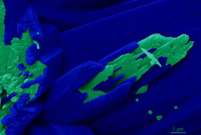
A high power microscopic image of the skeleton of Turbinaria peltata shows a model of ion attachment (in blue) and of nanoparticle attachment (in green) of new minerals to the skeleton, indicating that both systems are used to build coral skeletons. Credit: Pupa Gilbert
Coral reefs are vibrant communities that are home to a quarter of all species in the ocean and are indirectly crucial for the survival of others. But they’re slowly dying – by some estimates, 30 to 50 percent of reefs have been lost – due to climate change.
In a new study, physicists at the University of Wisconsin-Madison observed corals forming reefs at the nanoscale and identified how they create their skeletons. Findings explain how corals resist ocean acidification caused by increased carbon dioxide levels and suggest that controlling water temperature, not acidity, is key to mitigating losses. and restore reefs.
“Coral reefs are currently threatened by climate change. It’s not in the future, it’s in the present, ”says Pupa Gilbert, UW-Madison professor of physics and lead author of the study. “How corals deposit their skeletons is fundamentally important in assessing and helping their survival.”
Reef-forming corals are marine animals that produce a hard skeleton made up of aragonite, a form of the mineral calcium carbonate. But how the skeletons grow has remained unclear. One model suggests that calcium and carbonate ions dissolved in the calcifying fluid of corals bind one by one in the crystalline aragonite of the growing skeleton. A different model, proposed by Gilbert and his colleagues in 2017 and based on a study of a species of coral, suggests instead that undissolved nanoparticles bind and then slowly crystallize.
In the first part of a new study, published on November 9 in the Proceedings of the National Academy of Sciences, Gilbert and his research team used a spectromicroscopy technique known as PEEM to probe the growing skeletons of five freshly harvested corals, including representatives of the four possible forms of reef-forming coral: branched, massive, encrusting and table. PEEM chemical maps of calcium spectra have enabled scientists to determine the organization of different forms of calcium carbonate at the nanoscale.
PEEM results showed amorphous nanoparticles present in coral tissue, on the growth surface, and in the region between tissue and skeleton, but never in the mature skeleton itself, supporting the nanoparticle attachment pattern. However, they also showed that although the growth edge is not densely filled with calcium carbonate, the mature skeleton is an outcome that does not support the nanoparticle attachment model.
“If you imagine a bunch of spheres, you can never fill the space completely; there is always space between the spheres, ”says Gilbert. “So this was the first indication that attachment of nanoparticles may not be the only method.”
The researchers then used a technique that measures the exposed internal area of porous materials. Large geological crystals of aragonite or calcite – formed by something that is not alive – are found to have about 100 times less area than the same amount of material made of nanoparticles. When they applied this method to corals, their skeletons gave roughly the same value as large crystals, not nanoparticles.
“Corals fill space as much as a single crystal of calcite or aragonite. So the attachment of ions and particles has to occur,” says Gilbert. “The two separate camps that advocate particles over ions are in fact both correct.”
This new understanding of the formation of the coral skeleton can only make sense if one last thing is true: that seawater is not in direct contact with the growing skeleton, as is commonly assumed. In fact, recent studies of calcifying coral fluid have found that it contains slightly higher concentrations of calcium and three times more bicarbonate ions than seawater, which supports the idea that the skeleton in growth is effectively isolated from seawater.
Instead, the researchers come up with a model where corals pump calcium and carbonate ions from seawater through coral tissue, which concentrates these minerals near the skeleton. Importantly, this control allows corals to regulate their internal ion concentrations, even as the oceans acidify due to increased levels of carbon dioxide.
“Until this work, people had assumed that there was contact between seawater and the growing skeleton. We have shown that the skeleton is completely separate from seawater, and this has immediate consequences, ”says Gilbert. “If there are coral reef remediation strategies, they shouldn’t focus on tackling ocean acidification, they should focus on tackling ocean warming. To save coral reefs, we need to lower the temperature, not raise the pH of the water. ”
Coral skeletons can resist the effects of ocean acidification
Chang-Yu Sun el al., “From Particle Attachment to Coral Skeletons Filling Space,” PNAS (2020). www.pnas.org/cgi/doi/10.1073/pnas.2012025117
Provided by the University of Wisconsin-Madison
Quote: Better Understanding of Coral Skeleton Growth Suggests Ways to Restore Reefs (2020, November 9) Retrieved November 10, 2020 from https://phys.org/news/2020-11-coral-skeleton-growth-ways -reefs.html
This document is subject to copyright. Other than fair use for study or private research, no part may be reproduced without written permission. The content is provided for information only.
[ad_2]
Source link