
[ad_1]

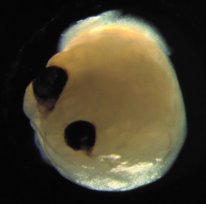
A brain organoid with optical sections. Credit: Elke Gabriel
Human-induced pluripotent stem cells (iPSCs) can be used to generate brain organoids containing an ocular structure called the optic slice, according to a study published Aug. 17 in the journal Stem cell. Organoids spontaneously developed bilaterally symmetrical optic sections from the front of the brain-like region, demonstrating the intrinsic self-structuring ability of iPSCs in a very complex biological process.
“Our work highlights the remarkable ability of brain organoids to generate primitive sensory structures sensitive to light and harboring cell types similar to those found in the body,” says lead study author Jay Gopalakrishnan of the Düsseldorf University Hospital. “These organoids can help study brain-eye interactions during embryonic development, model congenital retinal disorders, and generate patient-specific retinal cell types for personalized drug testing and transplant therapies.”
Many aspects of human brain development and disease can be studied using 3D brain organoids derived from pluripotent stem cells, which can give rise to all types of cells in the body. Researchers previously used human embryonic stem cells to generate the optical cut, which gives rise to the retina, the light-sensitive layer of tissue at the back of the eye. Another study demonstrated that optical cup-shaped structures can be generated from iPSC, which are derived from adult cells that have been genetically reprogrammed into an embryonic-like pluripotent state.

This graphic summary shows how the brain organoids of the optic vesicles are developed. Credit: Gabriel et al./Cell Stem Cell
In the past, the production of optic wells from pluripotent stem cells focused on the generation of the pure retina. Until now, optic cups and other 3D retinal structures have not been functionally integrated into brain organoids.
To achieve this feat, Gopalakrishnan and his team modified a protocol they had previously developed to transform iPSCs into neural tissue. Organoids in the human brain formed optical sections, which appeared as early as 30 days and became visible structures within 50 days. This period corresponds to that of the development of the retina in the human embryo and may make certain types of developmental neurobiology experiments more effective.
Out of 16 independent batches from four iPSC donors, the researchers generated 314 brain organoids, 72% of which formed optical sections, showing that the method is reproducible. These structures contained various types of retinal cells, which formed electrically active neural networks that responded to light. Brain organoids in the optic section also contained lens and corneal tissue and exhibited retinal connectivity with regions of the brain. “In the mammalian brain, nerve fibers from retinal ganglion cells connect to their brain targets, an aspect that has never been demonstrated before in an in vitro system,” explains Gopalakrishnan.
In future studies, they plan to develop strategies to keep the optic cups viable for long periods of time, using them to study the mechanisms that cause retinal damage.
Researchers use arched organoids to study hypothalamus development and disease
Elke Gabriel et al, Organoids of the Human Brain Assemble Functionally Integrated Bilateral Optic Vesicles, Stem cell (2021). DOI: 10.1016 / j.stem.2021.07.010
Quote: Brain Organoids Develop Optic Sections That Respond to Light (2021, August 17) retrieved August 18, 2021 from https://medicalxpress.com/news/2021-08-brain-organoids-optic-cups. html
This document is subject to copyright. Other than fair use for private study or research purposes, no part may be reproduced without written permission. The content is provided for information only.
[ad_2]
Source link