
[ad_1]

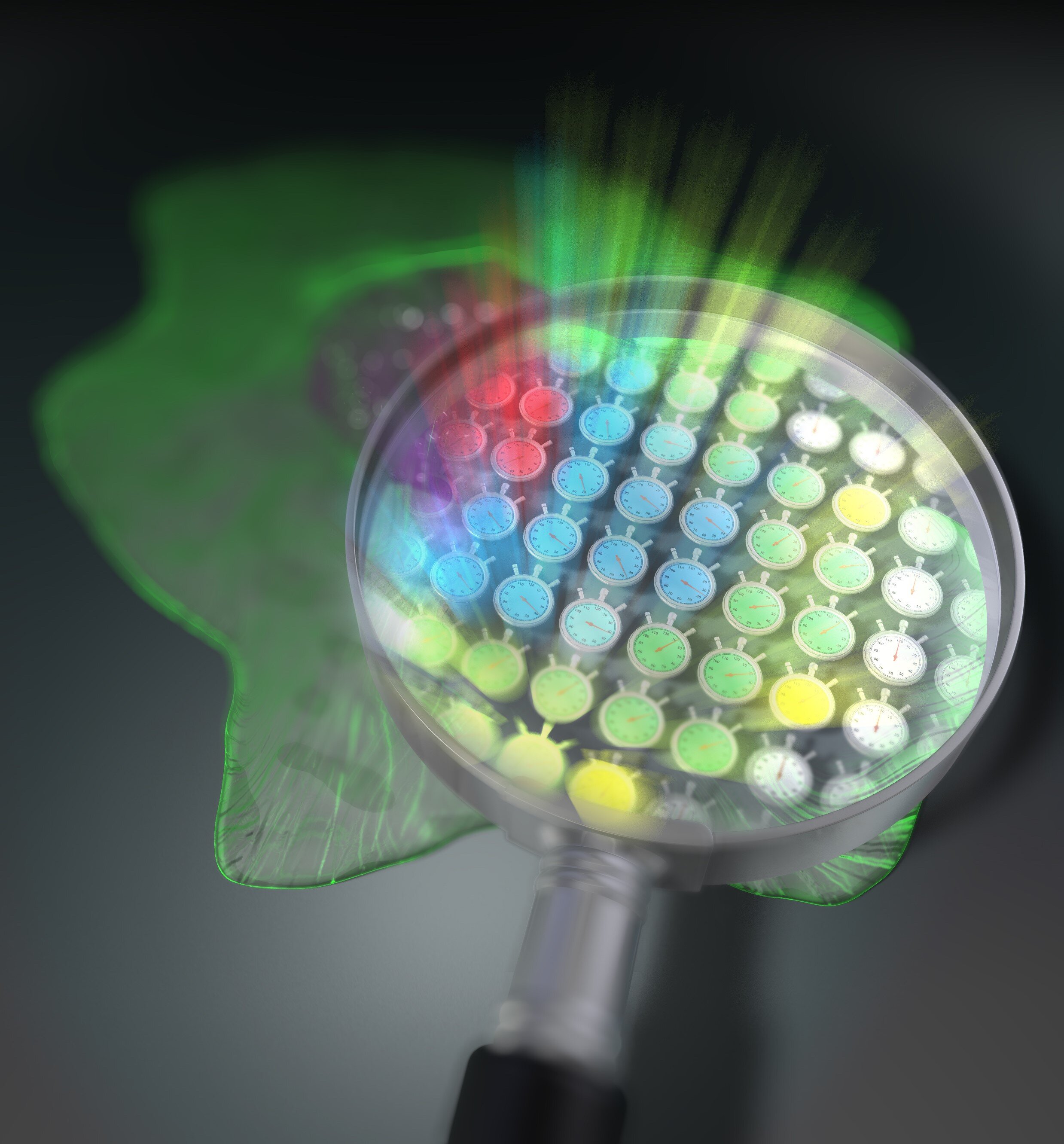
The 2D layout of 44,400 luminous chronometers enables lifetime fluorescence imaging without scanning. Credit: Tokushima University
Fluorescence microscopy is widely used in biochemistry and life sciences because it allows scientists to directly observe cells and certain compounds in and around them. Fluorescent molecules absorb light in a specific wavelength range and then re-emit it in the longer wavelength range. However, the main limitation of conventional fluorescence microscopy techniques is that the results are very difficult to assess quantitatively; the fluorescence intensity is significantly affected by the experimental conditions and the concentration of the fluorescent substance. Today, a new study by Japanese scientists is set to revolutionize the field of fluorescence lifetime microscopy.
One way to get around the conventional problem is to focus on the lifetime of the fluorescence rather than the intensity. When a fluorescent substance is irradiated with a short burst of light, the resulting fluorescence does not immediately disappear but “decays” over time in a way that is specific to that substance. The fluorescence lifetime microscopy technique exploits this phenomenon, independent of experimental conditions, to quantify fluorescent molecules and changes in their environment. However, the fluorescence decay is extremely fast and ordinary cameras cannot capture it. Although a single point photodetector can be used instead, it must be scanned across the entire sample area to be able to reconstruct a full 2D image from each measured point. This process involves the movement of mechanical parts, which greatly limits the speed of image capture.
In this recent study, published in Scientific advances, the team of scientists has developed a new approach to acquire lifetime fluorescence images without the need for mechanical scanning. Professor Takeshi Yasui, Institute of Post-LED Photonics (pLED), University of Tokushima, Japan, who led the study, said, “Our method can be interpreted as simultaneously mapping 44,400 ‘stopwatches’ light-based measure fluorescence lifetimes – all in one shot and without scanning. ”

This new fluorescence microscopy technique will measure both the intensity and the lifetime of fluorescence and will not require mechanical scanning of a focal point; instead, it will produce images of all points in the sample simultaneously, allowing a more quantitative study of dynamic biological and chemical processes. Credit: Suana Science YMY
One of the main pillars of their method is the use of an optical frequency comb as the excitation light for the sample. An optical frequency comb is basically a light signal made up of the sum of many discrete optical frequencies with constant spacing between them. The word “comb” in this context refers to the appearance of the signal when plotted against optical frequency: a dense cluster of equidistant spikes rising up from the optical frequency axis and resembling a comb. hair. Using special optical equipment, a pair of excitation frequency comb signals are broken down into individual optical beat signals (double comb optical beats) with different intensity modulation frequencies, each carrying a frequency single modulation and irradiated on the target sample. The key here is that each light beam hits the sample in a spatially distinct location, creating a one-to-one correspondence between each point on the sample’s 2D surface (pixel) and each modulation frequency of the double-comb optics. . Beats.
Due to its fluorescence properties, the sample re-emits part of the captured radiation while preserving the frequency-position correspondence. The fluorescence emitted by the sample is then simply focused using a lens on a high speed single point photodetector. Finally, the measured signal is transformed mathematically in the frequency domain, and the fluorescence lifetime at each “pixel” is easily calculated from the relative phase delay which exists between the excitation signal at this modulation frequency and that measured.
Thanks to its superior speed and high spatial resolution, the microscopy method developed in this study will make it easier to exploit the advantages of fluorescence lifetime measurements. “Because our technique does not require scanning, a simultaneous measurement on the whole sample is guaranteed with each take”, explains Professor Yasui, “This will be useful in the life sciences where dynamic observations of cells alive are necessary. In addition to providing a deeper insight into biological processes, this new approach could be used for simultaneous imaging of multiple samples for antigen testing, which is already in use for the diagnosis of COVID-19.
Perhaps more importantly, this study shows how optical frequency combs, which were only used as “frequency rulers”, can find a place in microscopy techniques to push the boundaries of life sciences. It holds promise for the development of new therapeutic options to treat intractable diseases and improve life expectancy, thereby benefiting all of humanity.
Scientists record wide spectra with nearly a hundred thousand colors in almost complete darkness
T. Mizuno et al. Lifetime full field fluorescence double comb microscopy using spectral mapping and double comb optical beat frequency multiplexing, Scientific advances (2021). DOI: 10.1126 / sciadv.abd2102, advances.sciencemag.org/content/7/1/eabd2102
Provided by Tokushima University
Quote: Comb of a life: A new method for fluorescence microscopy (2021, January 4) retrieved January 4, 2021 from https://phys.org/news/2021-01-lifetime-method-fluorescence-microscopy.html
This document is subject to copyright. Apart from any fair use for study or private research, no part may be reproduced without written permission. The content is provided for information only.
[ad_2]
Source link