[ad_1]
Neurosurgeons of the Clavel Institute, at the QuironSalud Hospital of Barcelona, performed lumbar surgery in the world with an 18-year-old Guatemalan who traveled to Spain for a double operation fracture in the fifth lumbar vertebra .
As reported by the health center, the intervention repaired a spondylolysis – a fracture – in the fifth lumbar vertebra without having to resort to the usual method, fixing the 39, injured joint, and avoid a reduction of mobility .
The intervention was performed on July 9 and was performed by doctors Pablo Clavel and Ignasi Català.
The patient, who was released four days after the operation, traveled specifically to Guatemala Spain to undergo this procedure after undergoing bilateral spondylolysis in the fifth lumbar vertebra since he was a teenager.
Spondylolysis is a fracture that causes a separation between the body of the vertebra – the anterior – and the posterior arch .
The fourth and fifth lumbar vertebrae are those who usually suffer most from this condition, which may have a conbad origin or be caused by bone consolidation problems during childhood or adolescence, or by trauma .
According to doctors at the Clavel Institute, spondylolysis may not present symptoms but often cause lumbalgias, and in some cases eventually drift into a spondylolisthesis, moving towards the front of the body of the fractured vertebra which causes more severe lumbago and sciatica.
New intervention
In the most severe cases of spondylolysis an arthrodesis is usually performed, which consists of attaching the damaged vertebrae to another by means of screws or grafts. bone and thus block the joint to achieve greater stability and less pain.
The novelty of this intervention is that it was not necessary to resort to this fixation, so the mobility of the spine did not
The surgeons, instead of performing an arthrodesis, fixing the fifth lumbar vertebra of the patient with his first sacral vertebra, opted for to repair the double fracture suffered by the young man.
Thus, joined the fractured bones with very small titanium screws and treated the bone with bone growth inducing protein BMP-2 . which helps to consolidate fractures
"Minimally Invasive"
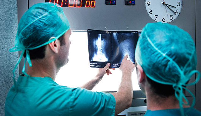
The intervention could be performed with minimally invasive surgery thanks to the # 39; use of an intraoperative n scanner which incorporates software allowing intraoperative navigation with high precision data, three-dimensional images during operations in the operating room and in real time.
Guided by the intraoperative CT scan, surgeons performed two small incisions in the patient's back and through tubular systems of progressive dilatation could approach and repair the vertebral fracture.
" The option of osteosynthesis was badessed as valid in this case because it was very patient young so there was more badurance that the fracture would be
"Thanks to this technique, the postoperative period is shorter." In less than a week, the patient is at home and, in two or three months, if the fracture is fully consolidated, he can return to normal life "they concluded.
Source link
