
[ad_1]
“African-American women continue to develop breast cancer at a younger age and often in more advanced stages,” Salewai Oseni, breast surgeon at Massachusetts General Hospital, said in a recent press release. “This, coupled with the higher case of triple negative breast cancer in this group, resulted in increased breast cancer mortality.”
Over the past two years, researchers at MIT CSAIL and the Abdul Latif Jameel Clinic for Machine Learning in Health have worked to develop a new deep learning system capable of predicting a patient’s cancer risk. by using only the person’s mammograms, which would work just as effectively regardless of race or ethnicity.
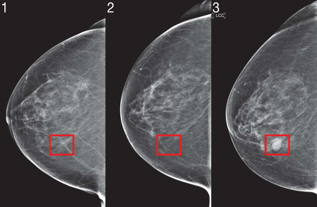
Nicknamed “Mirai” (not to be confused with Toyota’s Fuel Cell EV), this algorithm would be able to model “a patient’s risk over several future time points”, while taking into account minor deviations, up to the the clinic’s brand of mammography machine uses, according to a Wednesday release from MIT. Its predictions can be optimized further if other clinical risk factors – such as age or family history – are available.
The CSAIL team initially trained Mirai on a dataset of 200,000 exams from Massachusetts General Hospital (MGH) before validating their predictive results on additional sets from the Karolinska Institute in Sweden and Chang Gung Memorial Hospital in Taiwan. . So far, the results are very encouraging with results suggesting that Mirai is “significantly more accurate”, according to the release, at predicting cancer risks in patients in all three data groups and able to correctly identify close to twice as many potential cancer cases among high-risk groups as the Tyrer-Cuzick diagnostic model currently used in the study.
To ensure that Mirai’s recommendations were consistent, the CSAIL team skewed the algorithm by running it through an adversarial network to differentiate which aspects of the mammogram are important and those caused by random environmental variances. minor (such as make / model of mammography machine).
“Improved breast cancer risk models allow targeted screening strategies that allow earlier detection and less screening damage than existing guidelines,” Adam Yala, senior author of CSAIL Scientific translational medicine study, said in a statement. “Our goal is to incorporate these advances into the standard of care.”
WITH
This could advance the state of oncology science. Modern mammograms still suffer from reliability issues, even now 60 years after the technology’s widespread adoption. Experts still disagree on how often women should be screened, with some favoring more aggressive strategies to detect cancerous growths as early as possible, while others advocate longer gaps between them. routine testing in order to lower medical costs for patients). Mirai will be used to help doctors determine which patients would benefit most (and most equally) from additional imaging and MRI based on both the mammogram image and other factors such as the age, genetics, family medical history, and breast tissue density.
“We know that MRI can detect cancer earlier than mammography and that earlier detection improves patient outcomes,” Yala explained. “But for patients at low risk for cancer, the risk of false positives may outweigh the benefits. With improved risk models, we can design more nuanced risk screening guidelines that offer more sensitive screening, like MRI, to patients who will develop cancer to achieve better outcomes while reducing unnecessary screening. and over-processing for the rest. “
Mirai also takes into account risk factors that may not appear in mammography imaging, such as patient age, hormone levels, and menopausal status. These factors are ingrained during the training phase, which allows the model to predict them based on the mammography image given, even if the clinician has not manually provided this information.
In the future, Mirai may find use in other medical applications to benefit the community. Although the system is not able to interpret a patient’s history of existing imaging results and incorporate them into their assessment, it can rely on the additional X-rays / MRIs provided to it at the future. The team also plans to integrate tomosynthesis techniques to further increase Mirai’s statistical ability. The CSAIL team also partnered with researchers at Emory University to further validate the model.
[ad_2]
Source link