
[ad_1]
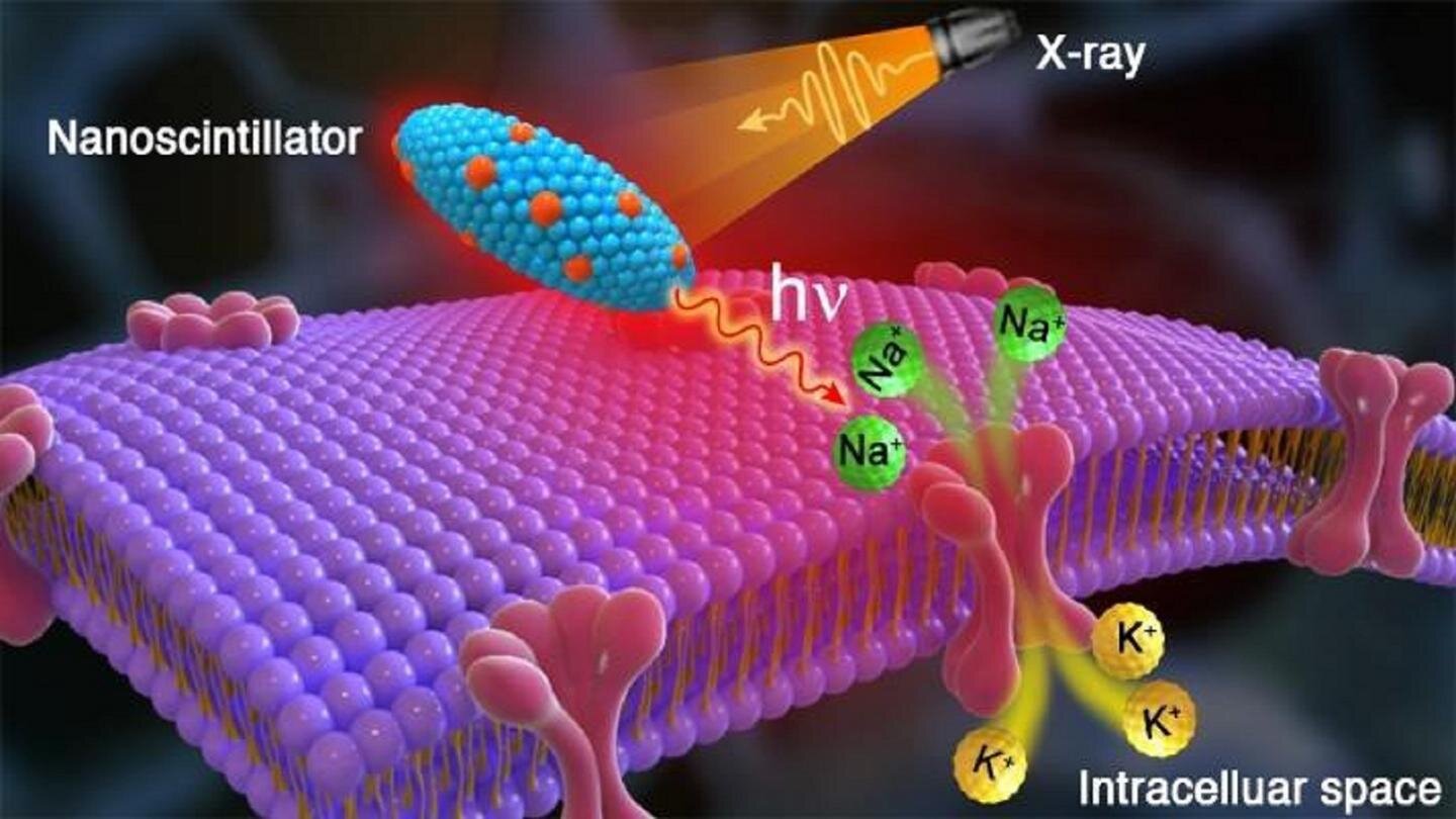
 and potassium (K +) ions and thus activates neurons in the brain. Credit: Zhaowei Chen / Argonne National Laboratory")
The artist’s rendering shows X-rays hitting radioluminescent nanoparticles in the brain, which emit red light that triggers an influx of sodium (Na +) and potassium (K +) ions and thus activates neurons in the brain. Credit: Zhaowei Chen / Argonne National Laboratory
Scientists make the crucial discovery of a method of wirelessly modulating neurons with X-rays that could improve the lives of patients with brain disorders. The x-ray source only requires a machine like the one found in a dentist’s office.
Many people around the world suffer from movement-related brain disorders. Epilepsy accounts for more than 50 million; essential tremor, 40 million; and Parkinson’s disease, 10 million.
Relief for some people with brain disorders may one day be underway in the form of a new treatment invented by researchers at the U.S. Department of Energy’s (DOE) Argonne National Laboratory and four universities. Treatment is based on breakthroughs in both optics and genetics. It would be applicable not only to movement-related brain disorders, but also chronic depression and pain.
This new treatment involves stimulating neurons deep in the brain with injected nanoparticles that light up when exposed to x-rays (nanoscintillators) and would eliminate invasive brain surgery currently underway.
“Our high-precision, non-invasive approach could become routine with the use of a small x-ray machine, the type commonly found in all dental offices,” said Elena Rozhkova, senior author and nanoscientist at the Center d’Argonne for nanoscale materials (CNM). , a facility for users of the DOE Science Office.
Traditional deep brain stimulation requires an invasive neurosurgical procedure for disorders when conventional drug therapy is not an option. In the traditional procedure, approved by the United States Food and Drug Administration, surgeons implant a calibrated pulse generator under the skin (similar to a pacemaker). They then connect it with an insulated extension cord to electrodes inserted into a specific area of the brain to stimulate surrounding neurons and regulate abnormal impulses.
“Hispanic-American scientist José Manuel Rodríguez Delgado demonstrated deep brain stimulation in an arena in the 1960s,” said Vassiliy Tsytsarev, University of Maryland neurobiologist and co-author of the study. “He immobilized a rabid bull charging at him by sending a radio signal to an implanted electrode.”
About 15 years ago, scientists introduced a revolutionary neuromodulation technology, “optogenetics,” which is based on the genetic modification of specific neurons in the brain. These neurons create a light-sensitive ion channel in the brain and, therefore, fire in response to external laser light. This approach, however, requires very fine fiber optic wires implanted in the brain and suffers from the limited depth of penetration of laser light through biological tissue.
The team’s alternative optogenetic approach uses nanoscintillators injected into the brain, bypassing implantable electrodes or fiber optic wires. Instead of lasers, they replace x-rays because of their greater ability to cross biological tissue barriers.
“The injected nanoparticles absorb X-ray energy and convert it into red light, which has a significantly greater penetration depth than blue light,” said Zhaowei Chen, former CNM postdoctoral fellow.
“Thus, the nanoparticles serve as an internal light source that allows our method to work without wires or electrodes,” Rozhkova added. Since the team’s approach can both stimulate and soothe small, targeted areas, Rozhkova noted, it has other applications besides brain disorders. For example, this could apply to heart problems and other damaged muscles.
One of the keys to the team’s success has been the collaboration between two of Argonne’s world-class facilities: CNM and Argonne’s Advanced Photon Source (APS), a user installation of the DOE Office of Science. Work on these facilities began with the synthesis and multi-tool characterization of nanoscintillators. In particular, the X-ray excited optical luminescence of the nanoparticle samples was determined at an APS beamline (20-BM). The results showed that the particles were extremely stable for months and upon repeated exposure to high intensity x-rays.
According to Zou Finfrock, a staff scientist at the APS 20-BM line and the Canadian light source, “they continued to shine a beautiful red-orange light.”
Next, Argonne sent nanoscintillators prepared by CNM to the University of Maryland for testing in mice. The University of Maryland team performed these tests for two months with a small, portable x-ray machine. The results proved that the procedure worked as expected. Mice whose brains had been genetically modified to respond to red light responded to x-ray pulses with brain waves recorded on an electroencephalogram.
Finally, the team from the University of Maryland sent the brains of the animals for characterization using X-ray fluorescence microscopy performed by scientists in Argonne. This analysis was carried out by Olga Antipova on the beamline of the microprobe (2-ID-E) at APS and by Zhonghou Cai on the hard X-ray nanoprobe (26-ID) operated jointly by CNM and APS.
This multi-instrument arrangement made it possible to see tiny particles residing in the complex environment of brain tissue with a super-resolution of tens of nanometers. It also made it possible to visualize neurons near and far from the injection site on a microscopic scale. The results proved that nanoscintillators are chemically and biologically stable. They do not move away from the injection site and do not degrade.
“Sample preparation is extremely important in these types of biological analyzes,” said Antipova, a physicist in the Division of X-ray Sciences (XSD) at APS. Antipova was helped by Qiaoling Jin and Xueli Liu, who prepared brain cuts only a few microns thick with jeweler’s precision.
“There is intense commercial interest in optogenetics for medical applications,” Rozhkova said. “Although still in the proof-of-concept stage, we expect our patent-pending wireless approach with small x-ray machines to have a bright future.”
The related article “Wireless optogenetic modulation of cortical neurons activated by radioluminescent nanoparticles” appeared in ACS Nano.
See schizophrenia: X-rays shed light on neuronal differences, point to treatment
Zhaowei Chen et al, Wireless optogenetic modulation of cortical neurons activated by radioluminescent nanoparticles, ACS Nano (2021). DOI: 10.1021 / acsnano.0c10436
Provided by the Argonne National Laboratory
Quote: New Method Could Shine ‘A Healing Light’ On The Brain For People With Movement Disorders (2021, March 24) Retrieved March 25, 2021 from https://phys.org/news/2021- 03-method-brain-movement-disorders .html
This document is subject to copyright. Other than fair use for private study or research purposes, no part may be reproduced without written permission. The content is provided for information only.
[ad_2]
Source link