
[ad_1]

Credit: Unsplash / CC0 Public Domain
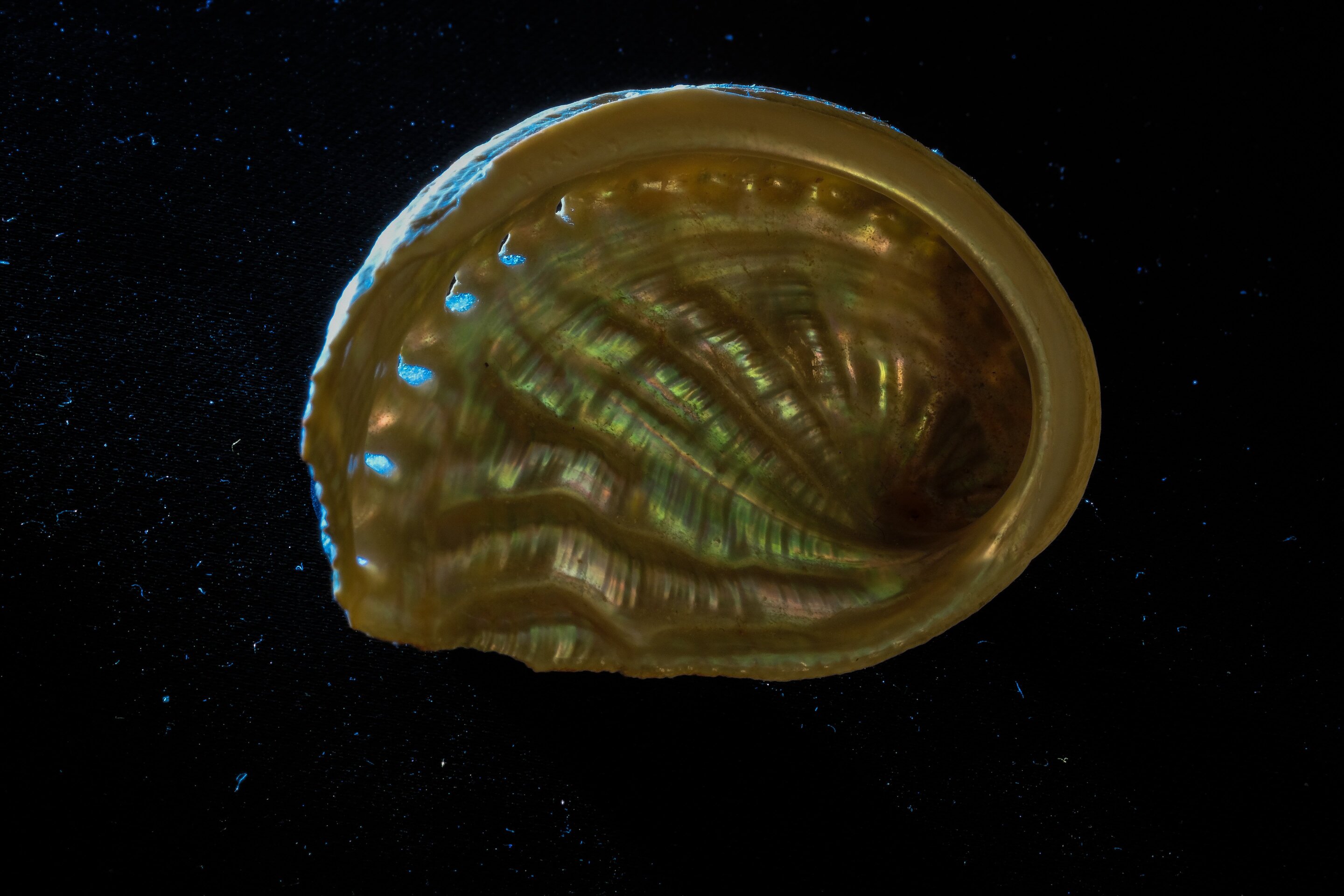
Most people are familiar with mother-of-pearl, an iridescent biomineral also known as mother-of-pearl, which comes from buttons, jewelry, instrument inlays, and other decorative ornaments. Scientists, too, have admired and marveled mother-of-pearl for decades, not only for its beauty and optical properties, but also for its exceptional toughness.
“It’s one of the most studied natural biomaterials,” says Pupa Gilbert, a professor of physics at the University of Wisconsin-Madison who has studied mother-of-pearl for more than a decade. “It may not look like much – just a shiny decorative material. But it may be 3,000 times more fracture resistant than aragonite, the mineral from which it is made. It has piqued the interest of materials scientists because the materials are better than the sum of their parts is extremely attractive. “
Now, a new non-destructive optical technique will unlock even more knowledge about mother-of-pearl and in doing so could lead to a new understanding of the history of climate. Gilbert, professor of electrical engineering at UW-Madison Mikhail Kats, their students and collaborators described the technique, called hyperspectral interference tomography, today in the journal Proceedings of the National Academy of Sciences.
Gilbert learned how mother-of-pearl forms, orders, resists fracture and how its layered structure registers the temperature at which it was formed. This mother-of-pearl layered structure reflects light and generates different colors depending on the thickness of the layer. This has led to an interest in finding a way to assess the thickness of the mother-of-pearl layers that does not involve destroying the mollusc shell in which it is deposited.
For help with this challenge, Gilbert turned to Kats and graduate student Jad Salman, who are experts in the study of optical phenomena.
For the project, Salman prepared 22 fresh red abalone shells for optical analysis. But taking optical spectra of mother-of-pearl is more difficult than it looks.
“If you want to probe this type of shell, which has a curved topography, it’s very difficult to get a good spectrum with a conventional spectrometer,” says Salman.
That’s why the team turned to a newer technology, hyperspectral photography, to image the full spectrum of the hull. From the start, they photographed the shells of industrial partner Middleton Spectral Vision before acquiring their own hyperspectral camera.
“It’s an imaging spectrometer where every pixel in the image gives a full spectrum,” says Salman. “When we use the camera in our setup, we can easily extract reliable spectral data over the large, uneven area of a shell in one shot.”
In addition to red abalone, the team also photographed mother-of-pearl from another species, the New Zealand paua shell, also known as rainbow abalone. Salman then used sophisticated modeling software he developed to determine the thickness of the mother-of-pearl layers pixel by pixel using the hyperspectral data.
The team calls the combination of hyperspectral interference tomography techniques and anticipates that it will be applicable to the measurement of other transparent layered structures found in plants, animals, geological samples or man-made materials.
For Gilbert, the new technique revealed a surprise about red abalone; he showed for the first time that the thickness of the mother-of-pearl layers thins as the mollusk ages. Because this thickness records the temperature of the seawater in which it is formed, the team believe it would be possible to use the technique to analyze fossil mollusc shells to learn more about past climates.
“This project is made up of a few different parts, each of which is fairly well understood,” Kats explains. “The power of this research is that we brought all this experimental and theoretical expertise, and were able to model not only well-designed and well-behaved layered structures, but also disordered and disordered biological structures. And we were able to get some useful information from that in a way that a biologist or paleoclimatologist can use. ”
New material mimics the strength and toughness of mother-of-pearl
Jad Salman et al., “Hyperspectral interference tomography of nacre”, PNAS (2021). www.pnas.org/cgi/doi/10.1073/pnas.2023623118
Provided by the University of Wisconsin-Madison
Quote: New non-destructive optical technique reveals mother-of-pearl structure (2021, March 29) retrieved March 29, 2021 from https://phys.org/news/2021-03-nondestructive-optical-technique-reveals-mother-of -pearl.html
This document is subject to copyright. Other than fair use for private study or research purposes, no part may be reproduced without written permission. The content is provided for information only.
[ad_2]
Source link