
[ad_1]
<div data-thumb = "https://scx1.b-cdn.net/csz/news/tmb/2019/engineeredpr.jpg" data-src = "https://scx2.b-cdn.net/gfx/ news / 2019 / engineeredpr.jpg "data-sub-html =" These magnetic protein crystals, isolated from cells, have been stained with a blue dye that binds to iron. Nano Letters 2019, DOI: 10.1021 / acs.nanolett.9b02266 ">
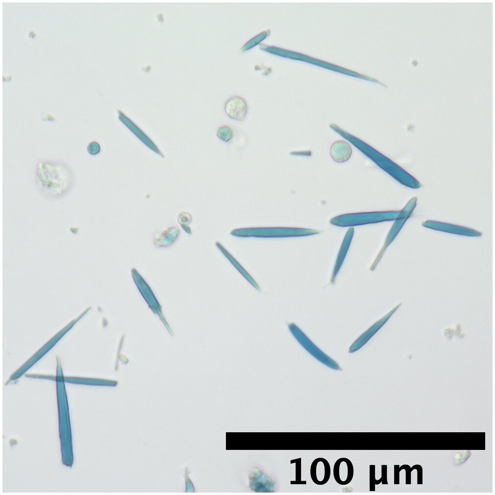
These magnetic protein crystals, isolated from cells, were stained with a blue dye that binds to iron. Credit: Adapted from Nano Letters 2019, DOI: 10.1021 / acs.nanolett.9b02266
If scientists could attribute magnetic properties to living cells, they might be able to manipulate cellular activities with external magnetic fields. But previous attempts to magnetize cells by producing iron-containing proteins had only resulted in weak magnetic forces. Now the researchers reporting in ACS Nano Letters have developed genetically encoded protein crystals capable of generating much stronger magnetic forces than those already reported.
The new field of magnetogenetics seeks to use genetically encoded, magnetic field-sensitive proteins to study and manipulate cells. Many previous approaches have put forward a natural iron storage protein called ferritin, which can self-assemble into a "cage" that can contain up to 4,500 iron atoms. Even with this large iron storage capacity, ferritin cages in cells generate magnetic forces millions of times too low for practical applications. To dramatically increase the amount of iron that a protein assembly can store, Bianxiao Cui and his colleagues wished to combine ferritin's ability to bond with iron with the self-assembling properties of one. another protein, called Inkabox-PAK4cat, that can form huge shaped crystals inside the cells. The researchers wondered whether they could cover the hollow interiors of ferritin protein crystals to store larger amounts of iron generating substantial magnetic forces.
To make the new crystals, the researchers fused genes encoding ferritin and Inkabox-PAK4cat and expressed the new protein in human cells in a petri dish. The resulting crystals, whose length reached about 45 microns (or about half the diameter of a human hair) after 3 days, did not affect cell survival. The researchers then opened the cells, isolated the crystals and added iron, which allowed them to attract the crystals with external magnets. Each crystal contained about five billion iron atoms and generated magnetic forces nine times higher than those of a single ferritin cage. By introducing preloaded iron crystals into living cells, the researchers were able to move them with a magnet. However, they were unable to magnetize the cells by adding iron to the crystals already growing in the cells, probably because the iron levels in the cells were too low. This is an area that requires further research, say the researchers.

Credit: American Chemical Society
What happens to magnetic nanoparticles in cells?
Thomas L. Li et al. Engineering of a genetically encoded magnetic protein crystal, Nano Letters (2019). DOI: 10.1021 / acs.nanolett.9b02266
Quote:
Artificial protein crystals make magnetic cells (September 25, 2019)
recovered on September 26, 2019
from https://phys.org/news/2019-09-protein-crystals-cells-magnetic.html
This document is subject to copyright. Apart from any fair use for study or private research purposes, no
part may be reproduced without written permission. Content is provided for information only.