.jpg)
[ad_1]
Researchers in the United States and Canada have described the structural basis and mode of action of two monoclonal antibodies that potently neutralize Severe Acute Respiratory Syndrome Coronavirus 2 (SARS-CoV-2) – the agent that causes the disease coronavirus 2019 (COVID-19).
Walther Mothes of Yale University School of Medicine in New Haven, Connecticut, and colleagues report that both antibodies – CV3-1 and CV3-25 – were effective against the SARS-CoV-2 variants of both concern. in vitro and in vivo.
The researchers say the antibodies were therefore prime candidates for elucidating the mode of action and identifying epitopes with pan-coronavirus activity.
“We believe that the two epitopes of these two antibodies are of interest for passive and active immunization strategies against emerging variants,” they write.
A pre-printed version of the research paper is available on the website bioRxiv* server, while the article is subject to peer review.
.jpg)
Emergence of SARS-CoV-2 variants threatens vaccination efforts
The SARS-CoV-2 virus is the third beta-coronavirus to have emerged in the human population since 2003 and is responsible for the COVID-19 pandemic which remains unchecked in many countries due to the emergence of new variants.
The viral spike protein, which mediates the initial stage of the infection process, is a primary target of the host’s immune response after natural infection. Therefore, this cutting-edge immunogen forms the basis of Moderna, Pfizer-BioNTech, Johnson & Johnson and AstraZeneca vaccines which are currently being deployed in many countries.
However, strains B.1.1.7 (alpha), B.1.351 (beta), P.1 (gamma) and B.1.617.2 (delta) all exhibit increased transmissibility and escape vaccine-induced immunity. and infection from previously circulating strains.
Learn more about the structure of the tip
The peak is made up of two subunits. The S1 subunit contains the receptor binding domain (RBD) which interacts with the host cell receptor angiotensin converting enzyme 2 (ACE2), while S2 contains the machinery required for host membrane fusion -viral after excretion of S1.
Cryo-electron microscopy (cryoEM) Cryo-electron tomography 70 (cryoET) has previously revealed several distinct pre-fusion conformations in which the RBD assumes “high” or “low” configurations. The ACE2 receptor binds to RBD and stabilizes it in the high configuration.
Antibodies isolated from convalescent or vaccinated individuals can be classified by their specificity for three main epitopes: RBD, the N-terminal domain (NTD) and the S2 subunit. For each class of antibody, conformational preference for RBD-up or RBD-down configurations has been described. Antibodies targeting RBD are often less potent against emerging variants of concern due to immune escape mutations that have evolved in this area.

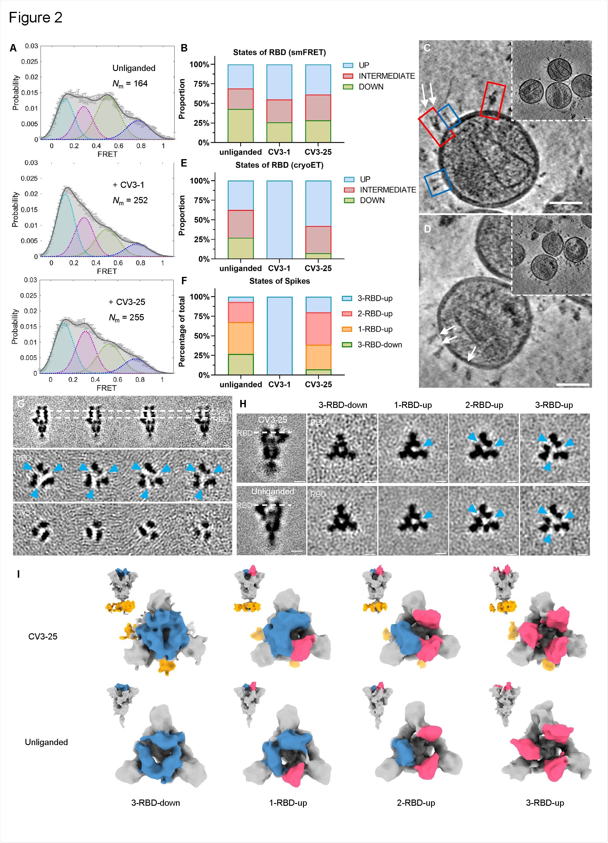
Conformational dynamics of SB.1.1.7 Linked with CV3-1 and CV3-25. (A) Conformational states of SB.1.1.7 on lentiviral particles monitored by smFRET for SB.1.1.7 without ligand, CV3-1 and CV3-25. FRET histograms with the number (Nm) of individual dynamic molecules / traces compiled into a conformation-population FRET histogram (gray lines) and fitted into a 4-state Gaussian distribution (solid black) centered on 0.1-FRET ( cyan dotted), 0.3- FRET (dotted red), 0.5-FRET (dotted green) and 0.8-FRET (dotted magenta). (B) Proportion of different RBD states identified by smFRET in (A). For comparative data parallel to cryoET data, the 0.8-FRET part has been omitted, due to its structural uncertainty. (CD) Enlarged views of S-carrying SARS-CoV-2 pseudoviruses linked by the CV3-1 (C) and CV3-25 (D) Fabs and representative slices of tomograms (inserts). Scale bar, 50 nm. White arrows indicate bound Fabs. Red boxes, prefusion tips; Blue boxes, post-fusion tips. (E) Proportion of different RBD states under different conditions from cyroET data. The UP state was separated by a classification targeted to the RBD region. The rest of the RBDs were defined as a DOWN state if there was no active RBD on the same peak, otherwise they were considered an INTERMEDIATE state. (F) Proportion of different peak RBD states on virions with and without bound Fab. The tips have been grouped into classes 3-RBD-down, 1-RBD-up, 2-RBD-up and 3-RBD-up. (G) Side views (top panel) and top views (middle and bottom panels) of the Averaged S subclasses linked by CV3-1 Fabs. (H) Side views (left column) of the consensus structure of unrelated S (bottom) and linked CV3-25 (top) and top views of subclass means (right columns) obtained after classification targeted at the RBD of S. In (GH), the dotted lines indicate the positions of the sections viewed from above. The blue arrowheads indicate the density difference between the RBD and the neighboring NTD that appears when the RBD enters the UP state. Scale bar, 5 nm. (I) Segmentation of the means of the subclasses of unrelated S (bottom) and CV3-25 (top) related. Top views and side views (insets) are shown for 3-RBD-down, 1-RBD-up, 2-RBD-up and 3-RBD-up classes. RBD down and RBD up are shown in blue and red respectively, Fab CV3-25 are shown in orange.
The potential of antibodies and cross-reacting vaccines
Although the immune responses induced by current vaccines offer varying degrees of protection against all known variants of concern, a booster injection may be necessary to provide sufficient protection against future variants.
In addition, given that SARS-CoV-2 is the third beta-coronavirus after SARS-CoV-1 and Middle East Respiratory Syndrome (MERS) to appear in humans this century and given the large natural reservoir of Similar viruses in wildlife such as bats, another coronavirus pandemic is likely to occur in the future.
The S2 domain of these coronaviruses is highly conserved, raising the possibility of cross-reacting antibodies and vaccines, explains Mothe and colleagues.
Researchers have previously characterized two potent spike binding antibodies – CV3-1 and CV3-25 – isolated from convalescent patients. While CV3-1 specifically targets RBD, CV3-25 recognizes the S2 domains of several beta-coronaviruses.
What was the current study?
First, the team tested the ability of antibodies to recognize and neutralize variants of concern B.1.1.7, B.1.351, P.1 and B.1.617.2 as well as B.1.429 (epsilon) , B Variants of interest .1.525 (eta), B.1.526 (iota) and B.1.617.1 (kappa).
The CV3-1 antibody efficiently bound to cells expressing the spike proteins of these different variants and exhibited potent neutralizing activity.
The CV3-25 antibody was less potent but still efficiently bound and neutralized all variants.
Both antibodies also limited viral replication in the nose and lungs of K18-hACE2 mice that received a lethal dose of the B.1.351 variant.
“These data demonstrate that unlike other antibodies which are attenuated against emerging variants, CV3-1 and CV3-25 remain potent against these variants and are therefore prime candidates to elucidate the mode of action and identify epitopes with pan-coronavirus activity. Wrote Mothes and his colleagues.
Study of the structure and mode of action of antibodies
Next, the researchers elucidated the structure and mode of action of the two antibodies by performing a viral-like particle (VLP) cryoET carrying the B.1.1.7 variant.
This revealed that CV3-1 bound to the 485-GFN-487 loop of RBD in the “high” conformation and induced a potent loss of S1.
“The data indicate that CV3-1 is a potent agonist and indicate that the 485-GFN-487 loop is a critical allosteric center for S1 activation,” the team explains.
In contrast, CV3-25 inhibited membrane fusion by binding to an epitope in the rod helix region of S2.
“Since the stem helix epitope is highly conserved among -coronaviruses, immunogens exhibiting this S2 epitope are attractive candidates for vaccines to cover all variants and possibly exhibit pan-coronavirus efficacy,” wrote Mothes and his colleagues.
What did the authors conclude?
The researchers claim that the epitopes of both antibodies are of interest for passive and active immunization strategies against emerging variants.
“Vaccine immunogen designs that incorporate the conserved regions in the RBD and the rod helix are candidates for eliciting protective immune responses against pan-coronavirus,” they conclude.
*Important Notice
bioRxiv publishes preliminary scientific reports which are not peer reviewed and, therefore, should not be considered conclusive, guide clinical practice / health-related behavior, or treated as established information.
[ad_2]
Source link