[ad_1]
Cardiac catheterization
involves inserting a catheter into the heart, through a femoral or aortic artery, to reach the coronary arteries, enter therapeutic equipment or for diagnostic purposes where the coronary arteries are injected with a Substance
Cardiac Catheterization:
Ask your doctor to perform a cardiac catheterization, a common medical procedure, to see if there is a heart problem or to correct a known heart problem. ] 1. Locate a narrow or blocked blood vessel
2. Evaluation of blood circulation
3- Diagnosis of the pumping function of cardiac blood, examination of the right and left ventricles, removal of cardiac tissue
4 Diagnosis of congenital heart defects present since birth
5.
Cardiac catheterization is also used in connection with certain procedures for the treatment of heart disease:
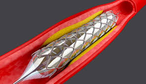
1 – at the time of installation of a Pillar
2 – Closure of Congenital Heart Holes
3. Expansion of vessels and treatment of leaking or narrowing of heart valves by repair or replacement.
4. Repair of an Industrial Valve Having Fled or Replaced an Industrial Valve
Risk of Cardiac Catheterization:
Cardiac catheterization rarely causes serious problems and some complications may occur, being susceptible to occur when older chronic kidney patients have diabetes.
1 – Bleeding, infection or pain at the site of insertion of the catheter
2 – Simple abrasion or hole in the blood vessels related to the heart
3. The allergic reaction to the dye used During of coronary angiography
4 arrhythmias, often go on their own, and recommend treatment if they continue.
5. Kidney damage caused by a dye used during coronary angiography
6. Blood clots Which can lead to a stroke, heart attack.
Nuclear Imaging:
While nuclear imaging is a type of imaging and medical examination using the amount
The patient radiates a dose of radiation by injection into the vein, and in cases taken orally, to deliver the radioactive material to the organ to be photographed without further.The amount, type and composition of the radioactive material vary according to the age of the patient and the member to photograph.
Nuclear imaging is diagnosed as a disease that can not be diagnosed by tests and laboratory techniques, in addition to its great utility in early diagnosis that helps to accelerate healing. 19659005] Nuclear medicine is used in the treatment and diagnosis of the mother
Tests in nuclear medicine:
1. Nuclear medicine establishes that tests do not require special preparation before being performed.
2. The patient is injected and placed under the gamma apparatus
3. The gamma camera transmits the radiation to an electrical current that travels to the camera's computer, This stream converts the information and accounts that appear on the computer's screen, and after having them analyzed and found results, printed images Patient film results
Nuclear Imaging Risk:
Simple quantities of radioactive material used in medically proven nuclear medicine trials that they do not affect the safety of the patient, so they are considered safe.
Examination and Radiation Protection Laws to Protect Patients and Workers
[ad_2]
Source link
