[ad_1]
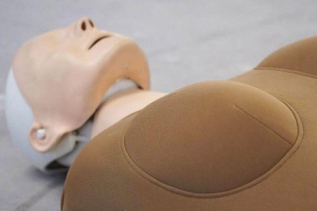
An advertising agency has created the very first attachment to convert a standard manikin into CPR into a female version.
The product, produced by the JOAN-based creative agency based in New York, was designed as a result of a recent study by Dr. Audrey Blewer that revealed that women suffered from cardiac arrest in are 27% less likely than men to benefit from CPR.
Research has determined that it's because people often do not know how to navigate CPR around a woman's breasts.
We will tell you what is true. You can form your own view.
Of
15p
$ 0.18
$ 0.18
$ 0.27
one day, more exclusive, analyzes and supplements.
As a result, JOAN has created a special attachment, called WoManikin, for standard dummy models, in order to allow trainees to get used to CPR on bodies with breasts.
In addition to addressing gender disparities, the advertising agency hopes that the model and her associated campaign will also help to overcome the discomfort felt by some men when it comes to CPR on a woman.
1/10 Subarachnoid vessels
Matt MacGregor Sharp, a PhD student at the University of Southampton, was a finalist. The very high resolution image shows a normal artery on the surface of the rat brain and was taken with a powerful scanning electron microscope. These "subarachnoid vessels" provide blood to the brain and also serve as a drain to eliminate toxic waste. Matt Macgregor's team is trying to show that the inability to remove waste from these vessels is one of the underlying causes of vascular dementia.
The researchers took the image using a technique called "freeze fracture" in which tissue or cell samples are frozen and then split to reveal the hidden layers within the sample so that they can be studied extremely detailed.
Seated above the brown brain tissue, the artery appears blue and its surrounding layer, the pia mater, is depicted in purple.
Matt MacGregor Sharp, University of Southampton, British Heart Foundation – Reflections of Research
2/10 Explosive debut
Winner: Endothelial cells line all blood vessels in the body, forming a barrier between circulating blood and the vessel wall. They also help protect the blood vessels from damage and release important chemical messengers that help control blood pressure.
Award-winning researcher Courtney Williams is a master's student and doctoral candidate at the University of Leeds. His lab is developing new methods for 3D mapping the growth of new blood vessels in their surrounding landscape. Understanding the complex secrets of blood vessel formation could be used to stimulate the regrowth of damaged blood vessels after a heart attack and to stop the growth of blood vessels when they are counterproductive.
Courtney Williams, University of Leeds, British Heart Foundation – Reflections on Research
3/10 A Snapshot of Platelet Production – Reflections from the Favorite of Research Supporters
This image of Abdullah Obaid Khan, a doctoral student at the University of Birmingham, won the fan favorite. What looks like precious jewelry is actually platelet formation in the bone marrow. Platelets are the smallest of our circulating blood cells and play an extremely important role in preventing bleeding. However, they also play a role in clot formation, which can lead to heart attacks and strokes. Abdullah Obaid Khan and his team are studying rare bleeding disorders.
Abdullah Obaid Khan, University of Birmingham, British Heart Foundation – Thoughts on Research
4/10 Cardiac Collagen Web Site – Shortlist
This colorful image shows the network resembling a network of the smallest blood vessels in the heart – the microvessels. Magenta marks the outer layer of collagen vessels; while orange marks their inner wall and blue the nuclei of the cells. Dr. Neil Dufton, Imperial College London
Dr. Neil Dufton, Imperial College London, British Heart Foundation – Thoughts on Research
5/10 Heart to Heart – Shortlist
This piece shows four ventricles (of a mouse) arranged in the form of four normal chambers hearts. The researchers used fluorescent markers to recognize certain proteins and created the image with the help of hundreds of images assembled. Dr. Elisa Avolio and Dr. Zexu Dang, University of Bristol
Dr. Elisa Avolio and Dr. Zexu Dang, University of Bristol, British Heart Foundation – Reflections of Research
6/10 Loving Artery – Selection
This image shows a cross section of an artery and the different layers that make up the wall of the artery. Silvia Lacchini, Affiliated Professor at the University of Glasgow
Silvia Lacchini, University of Glasgow, British Heart Foundation – Thoughts on Research
7/10 Oxidizing Ink Spot – Restricted List
This explosion of color shows one of the causes of cardiovascular disease – an enzyme called NADPH oxidase. The enzyme is considered "against Janus" because it is important for health as well as for diseases. This image shows the active enzyme in hypertensive patients. Dr. Livia de Lucca Camargo, University of Glasgow
Dr. Livia de Lucca Camargo, University of Glasgow, British Heart Foundation – Reflections on Research
8/10 Neon Skeleton – Shortlist
This image shows the developing blood vessel system of a two-day old zebrafish embryo. Researchers have used gene activators (on-off gene switches) to activate fluorescent markers in different types of endothelial cells – the important cells that line all blood vessels. All blood vessels activate the red marker, while the veins also activate the green marker, resulting in yellow veins and red arteries. Dr. Svanhild Nornes, University of Oxford
Dr. Svanhild Nornes, Oxford University, British Heart Foundation – Reflections on Research
9/10 Calcium Reef – Shortlist
This image shows the presence of calcium in the cells of the blood vessels of people with hypertension and resembling the reef of the Great Barrier Reef in Australia. Dr. Rheure Alves-Lopes, University of Glasgow
Dr. Rheure Alves-Lopes, University of Glasgow, British Heart Foundation – Reflections on Research
10/10 Blood vessels in the bud – Short list
This image shows the growing blood vessels in the mouse retina. In red, you can see all the blood vessels and in yellow / green you can see the actively growing blood vessels (process called germination). PhD student Kira Chouliaras, University of Oxford
Kira Chouliaras, University of Oxford, British Heart Foundation – Reflections on Research
This follows a 2018 survey by the University of Colorado that men are twice as likely as women to report fear of being accused of inappropriate touching or sexual assault as a reason for not having sex. not administer this life-saving act.
"JOAN's philosophy is based on a deep commitment to gender equality," said Jaime Robinson, co-founder and chief creative officer of JOAN.
"When we saw the study and this long-standing problem in the world of CPR, we found a relatively simple way to help make a difference.
"CPR models are designed to look like human bodies, but they actually represent less than half of our society," she added.
"The absence of women's corps in CPR training has the effect of making viewers hesitate, which results in women being more likely to die as a result of cardiac arrest. We hope WoManikin will bridge this education gap and, ultimately, save many lives. "
JOAN unveiled the product and launched its campaign to coincide with National CPR Awareness Week, which will take place June 1-7 in the United States.
The campaign includes a social media challenge on Instagram that asks women to share video clips of themselves with two-handed emojis on their hearts, including the hashtag #GiveMeCPR.
Support freethinking journalism and subscribe to Independent Minds
The British Heart Foundation (BHF) states that anyone witnessing a cardiac arrest must dial 999 and immediately begin CPR.
You can find out how to perform CPR and watch the BHF training video on CPR for a detailed demonstration here.
[ad_2]
Source link