
[ad_1]
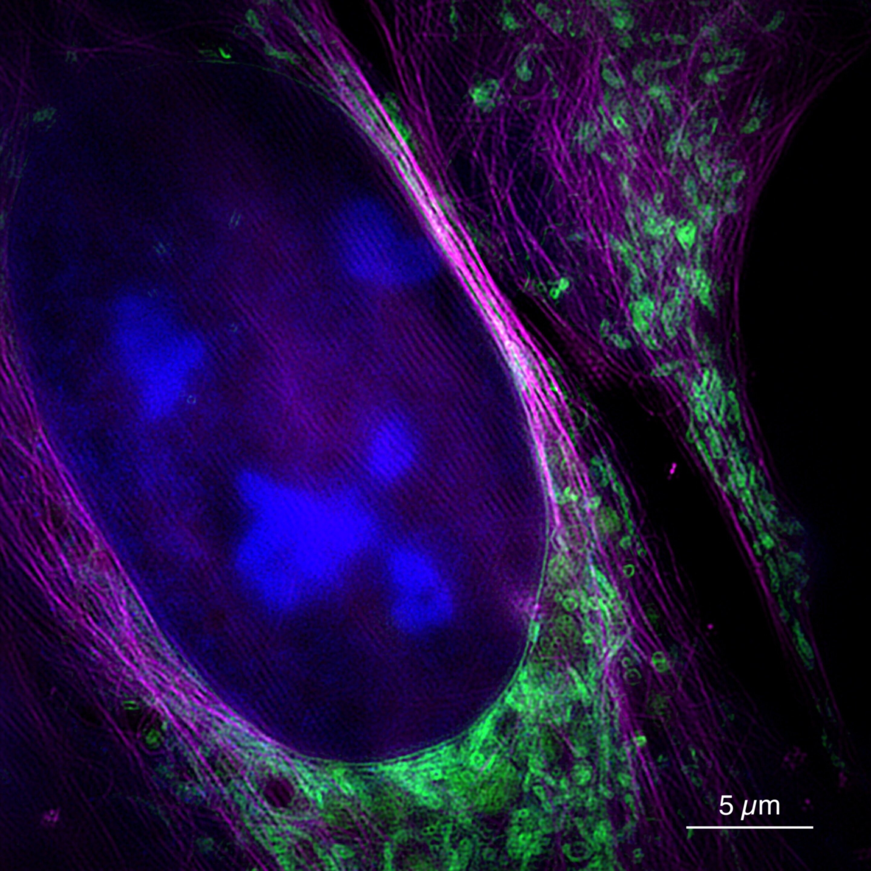
, mitochondria (in green) and a cytoskeleton (in magenta). Credit: University of Bielefeld / W. Hübner")
This image taken at the new microscope shows a living bone cancer cell with a nucleus (in blue), mitochondria (in green) and a cytoskeleton (in magenta). Credit: University of Bielefeld / W. Hübner
They can make tiny cell structures visible: microscopes at the tip of light offer resolutions of a few tenths of a nanometer, one millionth of a millimeter. Until now, super-resolution microscopes were much slower than conventional methods because more or less detailed data had to be recorded. Researchers from "Bielefeld" University and Jena partners developed the SR-SIM super resolution process. Academics have shown that SR-SIM is also possible in real time and at a very high imaging rate – and is therefore suitable for observing movements of very small cellular particles, for example. Their findings were published today (September 20) in the journal Nature Communications.
"This is what makes this type of microscopy very useful for applications in biology or medicine.The problem lies in the fact that microscopes with sufficiently high resolution can not display the information at the corresponding speed," says Professor Thomas Huser, Head of Biomolecular Research. Working Group on Physics at the University of Bielefeld. The SR-SIM project is funded by the German Research Foundation (DFG) and the European Union through the Marie Skłodowska-Curie Actions.
SR-SIM stands for "super-resolution structured illumination microscopy" and is a fluorescence microscopy procedure. The objects are irradiated with a laser light. This light excites special fluorescent molecules in the sample, so that they re-emit light at a different wavelength. The microscopic image then shows the re-emitted light. "Unlike other conventional methods of fluorescence microscopy, SR-SIM does not evenly illuminate samples, but with a fine, grid-like pattern.This special technology allows much higher resolution," Huser says. .
The procedure consists of two steps: the light re-emitted by the specimen is first recorded in several individual images. The final image is then reconstructed on a computer from these raw data. "The second stage, in particular, has cost a lot of time up to now," said Andreas Markwirth, also a member of the Bielefeld University's Biomolecular Physics Task Force and lead author of the 39; study. The researchers from Bielefeld have therefore worked with Professor Rainer Heintzmann of the Leibniz Institute for Photonic Technologies and the Friedrich Schiller University of Jena to speed up the process. The microscope is now designed to generate raw data more quickly. In addition, reconstruction of images takes much less time through the use of parallel computing on modern graphics cards.
For their study, the researchers tested the new method on biological cells and recorded motions of mitochondria, cell organelles of about a micrometer. "We have been able to produce about 60 frames per second, which is faster than movie movies, and the time between the measurement and the image is less than 250 milliseconds. real-time recording, "says Markwirth.
Until now, super-resolution methods have often been combined with conventional methods: a conventional fast microscope is used to search structures first. These structures can then be examined in detail with the help of a super-resolution microscope. "However, some structures are so small that they can not be found with conventional microscopes, for example specific pores in liver cells.Our method is both high resolution and fast, allowing biologists to explore such structures, "says Huser. Another application of the new microscope is the study of viral particles passing through the cell. "This allows us to understand exactly what happens during the infection process," says Huser. He expects the microscope to be used for such studies at the University of Bielefeld over the next year.
Super-resolution microscopes have only been around for about 20 years. In 1873, Ernst Abbe had discovered that the resolution of an optical system for visible light was limited to about 250 nanometers. In recent years, however, several optical methods have been developed to break what is now Abbé's diffraction barrier. In 2014, William E. Moerner and Eric Betzig, both from the United States, and Stefan Hell from Germany, received the Nobel Prize in Chemistry for developing a super-resolution of 20 to 30 nanometers .
New open source software for high resolution microscopy
Andreas Markwirth et al. Multicolored structured video illumination microscopy with real-time simultaneous reconstruction Nature Communications (2019). DOI: 10.1038 / s41467-019-12165-x
Quote:
HD microscopy in milliseconds (September 20, 2019)
recovered on September 20, 2019
at https://phys.org/news/2019-09-hd-microscopy-milliseconds.html
This document is subject to copyright. Apart from any fair use for study or private research purposes, no
part may be reproduced without written permission. Content is provided for information only.
[ad_2]
Source link