
[ad_1]
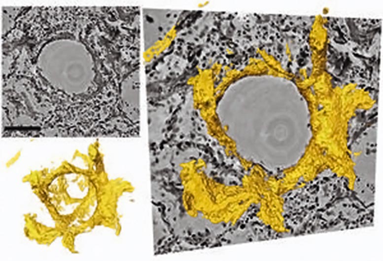
Sections through the three-dimensional reconstruction volume (top left, gray) around a hyaline membrane lung alveolus (bottom left, yellow). On the right, the images are superimposed. In the center is the air bubble (alveolus). The electron density is represented by different shades of gray. Inside the air bubble is a layer of protein and dead cell residue, the “hyaline membrane”. This deposit, which can be represented in its three-dimensional structure for the first time by the new method, reduces gas exchange and leads to respiratory distress. Credits: T Salditt, M Eckermann
Researchers led by The University of Göttingen is developing a new three-dimensional imaging technique to visualize tissue damage in severe Covid-19.
Physicists from the University of Göttingen, together with pathologists and lung specialists from the Medical University of Hanover, have developed a three-dimensional imaging technique that enables high-resolution, three-dimensional representation of damaged lung tissue following a severe Covid-19. Using a special x-ray microscopy technique, they were able to visualize the changes caused by the coronavirus in the structure of the alveoli (the tiny air sacs in the lungs) and the vascular system. The results of the study were published in the research journal eLife.
In severe disease of Covid-19, researchers observed significant changes in the vascular system, inflammation, blood clots, and “hyaline membranes”, which are made up of proteins and dead cells deposited on the alveolar walls, which makes gas exchange difficult or even impossible. With their new imaging approach, these changes can be visualized for the first time in larger tissue volumes, without cutting and staining or damaging the tissue as in conventional histology. It is particularly well suited for tracing small blood vessels and their branches in three dimensions, locating cells of the immune system that are recruited at sites of inflammation and measuring the thickness of the alveolar walls. Due to the three-dimensional reconstruction, the data could also be used to simulate gas exchange.
Professor Tim Salditt. Credit: University of Göttingen
“Using zoom tomography, large areas of wax-encrusted lung tissue can be scanned, allowing detailed examination to locate areas of particular interest around inflammation, blood vessels or bronchial tubes.” , explains lead author Professor Tim Salditt of the Institute for X-ray Physics at the University of Göttingen. Since x-rays penetrate deep into tissue, it allows scientists to understand the relationship between the microscopic structure of tissue and the larger functional architecture of an organ. This is important, for example, to visualize the tree of blood vessels down to the smallest capillaries.
The authors predict that this new radiography technique will be an extension of traditional histology and histopathology, areas of study that date back to the 19e century when optical microscopes had just become available and pathologists could thus disentangle the microscopic origins of many diseases. Even today, pathologists still follow the same basic steps to prepare and study tissue: chemical fixation, slicing, staining, and microscopy. This traditional approach, however, is not sufficient if three-dimensional images are required or if large volumes have to be projected, digitized or analyzed with computer programs.
Three-dimensional imaging is well known from computerized medical tomography (CT). However, the resolution and contrast of this conventional technique are not sufficient to detect tissue structure with cellular or subcellular resolution. Therefore, the authors used “phase contrast”, which exploits the different propagation speeds of X-rays in tissues to generate an intensity diagram on the detector. Salditt and his research group at the Institute for X-Ray Physics developed special optics and lighting algorithms to reconstruct sharp images from these models, an approach they have now adapted for studying the lung tissue affected by severe progression of Covid-19. Göttingen’s team could record lung tissue at scalable size and resolution, giving both larger insights and close-up reconstructions. Depending on the setting, their method can even give structural details lower than the resolution of conventional light microscopy. To achieve this, the researchers used very powerful X-radiation generated on the PETRAIII storage ring of the German electronic synchrotron (DESY) in Hamburg.
As was the case when the modern microscope was invented 150 years ago, significant advances have resulted from collaboration between physicists and medical researchers. The interdisciplinary research team hopes that the new method will support the development of treatment methods, drugs to prevent or mitigate severe lung damage from Covid-19, or to promote regeneration and recovery. “Only when we can clearly see and understand what is really going on, can we develop targeted interventions and drugs,” adds Danny Jonigk (Medical University of Hanover), who led the medical part of the interdisciplinary study.
Reference: “3D virtual pathohistology of lung tissue from COVID-19[feminine patients basés sur la tomographie à rayons X à contraste de phase »par Marina Eckermann, Jasper Frohn, Marius Reichardt, Markus Osterhoff, Michael Sprung, Fabian Westermeier, Alexandar Tzankov, Mark Kühnel, Danny Jonigk et Tim Salditt, 20 août 2020, eLife.
DOI: 10.7554 / eLife.60408
medRxiv
[ad_2]
Source link