[ad_1]
After 10 years of research and development, a company unveiled the first-ever X-ray color scanner
The boring old black and white. White X-rays are a thing of the past – after 10 years of development, MARS Bioimaging has unveiled the world's first color X-ray scanner. The device offers physicians an unprecedented tool for scanning patients' bodies, with potential applications ranging from research to diagnosis.
Color me surprised
X-ray imaging works by pushing high energy radiation into a plate recording (or "film"). The denser pieces inside you, especially bones, block or absorb these rays. Your most fleshy pieces – such as muscles, organs, and other soft tissues – usually allow X-rays to pass straight through. The radiation enters a particular area, exits the body and reacts with the recording plate. The soft tissues, which let a lot of this radiation pass, will appear dark on the recording. Areas that have allowed relatively little radiation to pass will appear white.
This is a really great way to look inside the body. It produces only black and white images and often with fuzzy outlines, but it's usually pretty accurate for what we need to do. A doctor can always know if one of your bones is broken by looking at an x-ray. It does not matter if the image shows only two colors without sharp lines because the bones tend to be pretty obvious.
With the new technology, the "X-ray" part of the new device works the same way. The real breakthrough is what she does with the readings
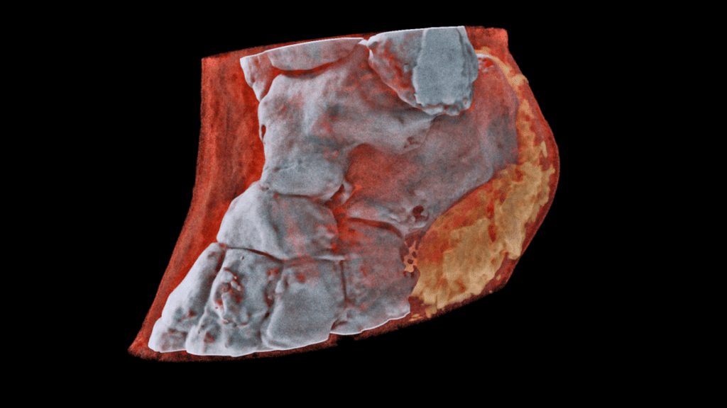
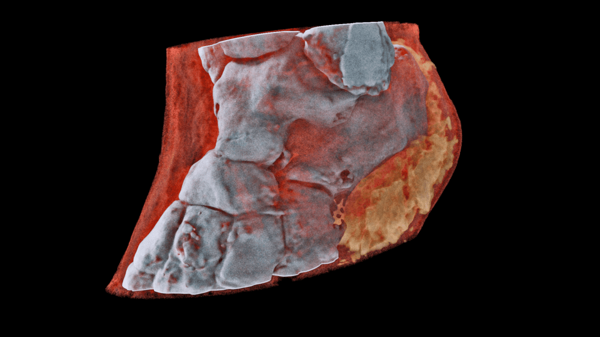
A human ankle seen through the new X-ray device.
Image credits: MARS Bioimaging.
Strictly speaking, the scanner itself does not produce the colors – they are generated after the readings are completed. The device uses a combination of Medipix technology. "Medipix is a family of chips read for imaging and particle detection," Cristina Agrigoroae wrote for CERN (European Organization for Nuclear Research). It works like a camera, detecting and counting every particle hitting the pixels when its electronic shutter is open.
Medipix was first developed to help CERN researchers track particles in the Large Hadron Collider – and computer algorithms for "measuring". Color Essentially, it all boils down to the way the device records radiation after it passes through a patient's body. Traditional X-ray machines record whether these waves pass through bone or soft tissue; Meanwhile, the MARS Bioimaging device records the intensity of outgoing radiation. Depending on these values, which depend on the composition of the tissues that they pass through, the algorithm fills the colors to represent bones, muscles and other tissues.

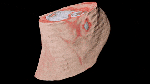
Timepix3, one of the texts read
Image credits CERN
But, if traditional X-rays are just good enough to spot fractures, why do we need all these colors? Are not they just going to confuse our doctors? Well, not really.
The main advantages of the new technology are a much better resolution (sharp images) and the ability to detect problems with bones and surrounding tissue.
"This technology distinguishes the machine from a diagnostic standpoint: its small pixels and precise energy resolution allow this new imaging tool to obtain images that no other tool can." imaging can not reach, "says Phil Butler, a physics professor, co-founder of MARS Bioimaging and one of the researchers behind the device.For example, Phil and his partner Anthony Butler (the son of Phil and a professor of bioengineering) have already made the device available for a number of studies in areas that had little use for X-ray scanners. "In all of these studies, Promising preliminary results suggest that when spectral imaging is commonly used in clinics, it will enable more accurate diagnosis and customization. Anthony Butler explains:
The duo plans to test their scanner in a New Zealand trial focused on patients in orthopedics and rheumatology. However, they warn that even if the lawsuit goes bad, it could still take years for the technology to get all the regulatory approval it needs before it can be used on a large scale.
Did you like this article? Join more than 40,000 subscribers to the ZME Science newsletter. Subscribe now!
Source link
