
[ad_1]
A high contrast black-and-white image of your bones is an effective tool for identifying fractures or fractures. But after more than 120 years, X-ray imaging gets a remarkable update with 3D color images that reveal far more than the bones inside you. These images will improve what a doctor can diagnose without opening you up.
The traditional approach to imaging the interior of a patient is to blast them with X-rays. This electromagnetic radiation has a shorter wavelength than visible light, so that it can easily pass through the soft tissues, but it has more difficulty in getting through harder materials like bones. On the other side of your body, a sensor or film produces an image based on the intensity of the X-rays that pass through it, revealing what is in you.
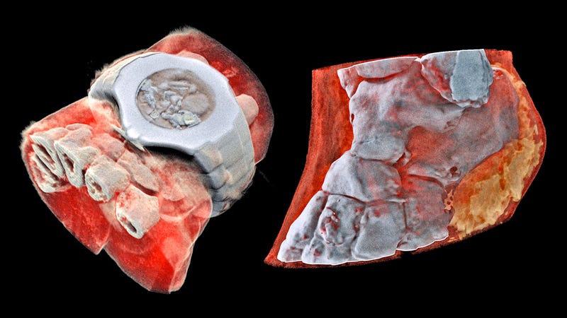
A New Zealand company called Mars Bioimaging has developed a new type of medical imaging scanner that works in the same way, but borrows the technology developed for CERN's Large Hadron Collider to produce results much more detailed. The Medipix3 chip works the same way as the sensor in your digital camera, but it detects and counts the particles that reach each pixel when a shutter opens.

When used in the new scanner developed by Phil and Anthony Butler of the Canterbury and Otago Universities in New Zealand, the Medipix3 chip, enhanced by algorithms Custom data processing, can detect the change in wavelength when x-rays pass through different materials in the body. This allows the scanner to differentiate bones, muscles, fats, liquids and all other materials from the human body, while other software uses this data to produce stunning color images that allow for three-dimensional vision of the human body. inside the body.
So, while a doctor examines the images of your arm, that he is looking for signs of fracture or fracture after an unpleasant fall, he may also look for other potentially dangerous medical conditions which might not appear in the usual X-rays. results. In fact, smaller trial versions of this scanner are already being used to study cancer, as well as bone and joint health in patients, but the technology will be useful in countless other medical fields, from dentistry to brain surgery.
It will be years before the new CT Spectral scanner receives all the approvals and approvals it needs for use in hospitals and clinics. But the stages of research are already well advanced and clinical trials are expected to begin in New Zealand in the coming months.
[Mars Bioimaging via CERN via New Atlas]
Source link
